. 2013 Mar 15;126(Pt 6):1429-39.
doi: 10.1242/jcs.118182. Epub 2013 Feb 1.

ERdj3 regulates BiP occupancy in living cells

Affiliations

ERdj3 regulates BiP occupancy in living cells

Feng Guo et al. J Cell Sci. .

Abstract

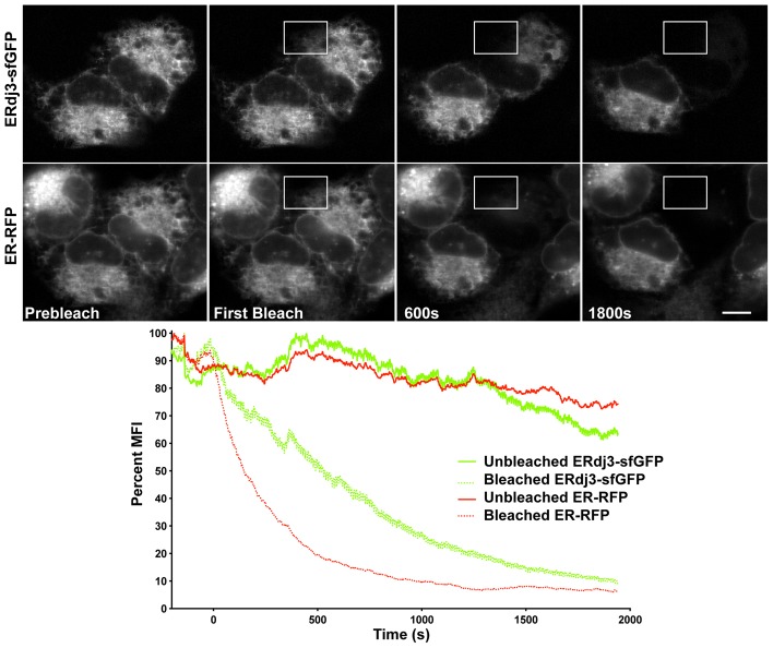
Co-chaperones regulate chaperone activities and are likely to impact a protein-folding environment as much as the chaperone itself. As co-chaperones are expressed substoichiometrically, the ability of co-chaperones to encounter a chaperone is crucial for chaperone activity. ERdj3, an abundant soluble endoplasmic reticulum (ER) co-chaperone of the Hsp70 BiP, stimulates the ATPase activity of BiP to increase BiP's affinity for client (or substrate) proteins. We investigated ERdj3 availability, how ERdj3 levels impact BiP availability, and the significance of J proteins for regulating BiP binding of clients in living cells. FRAP analysis revealed that overexpressed ERdj3-sfGFP dramatically decreases BiP-GFP mobility in a client-dependent manner. By contrast, ERdj3-GFP mobility remains low regardless of client protein levels. Native gels and co-immunoprecipitations established that ERdj3 associates with a large complex including Sec61α. Translocon binding probably ensures rapid encounters between emerging nascent peptides and stimulates BiP activity in the crucial early stages of secretory protein folding. Importantly, mutant BiP exhibited significantly increased mobility when it could not interact with any ERdjs. Thus, ERdjs appear to play the dual roles of increasing BiP affinity for clients and regulating delivery of clients to BiP. Our data suggest that BiP engagement of clients is enhanced in ER subdomains enriched in ERdj proteins.

Keywords: BiP; ERdj; FRAP; Fluorescence recovery after photobleaching; Quality control; Superfolder GFP.


Figures

Fig. 1.
Fig. 1.
Construction and characterization of ERdj3-sfGFP. (A) Illustration of fusion of human ERdj3 in-frame to sfGFP. (B) ERdj3-sfGFP colocalizes (yellow in merge panel) with ER-RFP in the ER of a co-transfected U2OS cell. Scale bar: 10 µm. (C) ERdj3-sfGFP is N-linked glycosylated in the ER. Portion of endogenous ERdj3 (lower band) in both untransfected (Un) and ERdj3-sfGFP transfected cells migrates faster (-CHO arrow) in an immunoblot of transiently transfected MDCK cell lysate stained with anti-ERdj3 when cells were treated with 1 µg/ml Tm for 8 hours. Similarly, ERdj3-sfGFP (upper band) migrates faster (-CHO arrow) when cells were treated with Tm. Molecular markers (kDa) are indicated.
Fig. 2.
Fig. 2.
ERdj3-sfGFP binds BiP and forms homo-oligomers. (A) ERdj3-sfGFP associates with BiP. Endogenous BiP is co-immunoprecipitated with anti-GFP when ERdj3-sfGFP is expressed in MDCK cells. Results are displayed in an immunoblot probed with anti-BiP (upper panel) or anti-GFP (lower panel). In the left lanes, transiently transfected MDCK cell lysates contain endogenous BiP bands [untransfected (Un), ERdj3-sfGFP (wt) or H53Q ERdj3-sfGFP (H53Q)]. Only the wild type binds BiP in the co-IP lanes. (B) ERdj3-sfGFP oligomerizes with ERdj3-mCherry. An immunoblot of cell lysates and immunoprecipitations was probed with anti-RFP (upper panel) or anti-GFP (lower panel). ERdj3-mCherry is pulled down with anti-GFP when coexpressed in MDCK cells with ERdj3-sfGFP, but ER-mCherry is not, demonstrating the interaction is specific for ERdj3. Asterisk (*) indicates a nonspecific band. (C) ERdj3-sfGFP oligomerizes with ERdj3-mCherry, but the F326D mutant ERdj3-sfGFP does not. Immunoblot probed with anti-RFP. ERdj3-mCherry is pulled down with anti-GFP when coexpressed in the MDCK cell with ERdj3-sfGFP, but not with F326D ERdj3-sfGFP. Molecular markers (kDa) are indicated.
Fig. 3.
Fig. 3.
ERdj3-sfGFP is mobile throughout the ER. U2OS cells, transiently co-transfected with ERdj3-sfGFP and ER-RFP, were subjected to dual color FLIP. Both ERdj3-sfGFP and ER-RFP fluorescence were homogenously depleted from the entire ER (upper panel). The graph below shows the rate of fluorescence depletion. ERdj3-sfGFP (green dotted line) fluorescence was depleted, but much more slowly than ER-RFP (red dotted line). Fluorescence loss was specific, as adjacent cells remained fluorescent (solid green and red lines). Scale bar: 10 µm.
Fig. 4.
Fig. 4.
Quantification of ERdj3-sfGFP mobility in the ER. (A) FRAP analysis of transiently transfected MDCK cells coexpressing ER-RFP and ERdj3-sfGFP. Images of cells after photobleaching the ROI (boxed area) reveal that ER-RFP recovers much more rapidly than ERdj3-sfGFP. Scale bar: 10 µm. (B) Representative fluorescence recovery plots illustrate the dramatically faster diffusion of ER-RFP relative to ERdj3-sfGFP. (C) Quantification reveals that ERdj3-sfGFP diffuses much more slowly than BiP-mGFP and ER-sfGFP. FRAP of MDCK cells transiently transfected with ERdj3-sfGFP, ER-sfGFP and BiP-mGFP. D values for single cells are plotted; horizontal lines indicate mean D values; P values are given above the columns.
Fig. 5.
Fig. 5.
The low mobility of ERdj3-sfGFP is independent of BiP binding, client levels and oligomerization. (A) Transiently transfected MDCK cells expressing ERdj3-sfGFP or the H53Q or F326D ERdj3-sfGFP mutants were analyzed by FRAP. (B) MDCK cells transiently transfected with ERdj3-sfGFP were treated with 0.2 µM Pact for 1 hour and analyzed by FRAP. (C) Transiently transfected MDCK cells expressing ERdj3-sfGFP, F326D ERdj3-sfGFP or V153A/F326D ERdj3-sfGFP mutants were analyzed by FRAP. D values for single cells are plotted; horizontal lines indicate mean D values; P values are given above the columns; error bars indicate s.e.m.
Fig. 6.
Fig. 6.
ERdj3-sfGFP and endogenous ERdj3 associate with a large complex in the ER. (A) Endogenous ERdj3 in untransfected (Un) MDCK cell lysate stained with anti-ERdj3 migrates significantly slower than expected on the immunoblot of a 7.5% tricine native gel. Transiently transfected ERdj3-sfGFP migrates similarly slowly in the anti-GFP immunoblot (8 or 12 hours). Slow migration of ERdj3-sfGFP appears to be independent of the expression level, as the migration pattern does not change at later times post transfection. (B) ERdj3 complex migrates distinctly more slowly than PDI and GRP94. U2OS cells were lysed in native lysis buffer and lysates were separated on a native gel, transferred to nitrocellulose and probed with the indicated antibodies. Note that GRP94 is an obligate dimer, consistent with its relatively slow migration. (C) ERdj3-sfGFP associates with the ER translocon Sec61 complex. ERdj3-sfGFP in transiently transfected MDCK cells associates with Sec61α in an anti-GFP IP. Immunoblots were probed with anti-Sec61α or anti-GFP. In the whole cell lysate lanes, both untransfected (Un) and ERdj3-sfGFP-expressing cell lysates contain bands corresponding to Sec61α. In the co-IP lanes, Sec61α is pulled down only in cells expressing ERdj3-sfGFP. Molecular markers are indicated in kDa.
Fig. 7.
Fig. 7.
Interactions with ERdj proteins regulate BiP availability in the ER lumen. (A) Transiently transfected MDCK cells coexpressing BiP-mCherry alone (Un) or with ERdj3-sfGFP (wt) or the BiP binding mutant H53Q ERdj3-sfGFP (H53Q) were analyzed by FRAP. (B) ERdj proteins regulate BiP availability in cells. FRAP analysis of MDCK cells transiently transfected with either wild-type or a mutant R197H BiP-mGFP, unable to bind ERdj proteins. Cells were untreated (un) or incubated with 1 µg/ml Tm for 4 hours. The R197H mutation significantly increased BiP mobility during both homeostasis and the ER stress. (C) The mobility of a BiP substrate-binding mutant (T453D) is resistant to ERdj3 overexpression in MDCK cells analyzed by FRAP. (D) ERdj3-sfGFP associates with a substrate-binding BiP mutant (T453D). Transiently transfected MDCK cells expressing the indicated proteins were lysed in IP buffer, incubated with anti-GFP beads and co-IPs, or whole cell lysate was separated by gel electrophoresis and probed with anti-GFP or anti-RFP, as indicated. Images are from the same film exposure of blot for a single gel. (E) Nascent substrate levels regulate the impact of ERdj3 on BiP diffusion in MDCK cells expressing BiP-GFP with or without pactamycin or ERdj3-mCherry and analyzed by FRAP. D values for single cells are plotted; horizontal lines indicate mean D values; P values are given above the columns; error bars indicate s.e.m.

Similar articles

Cited by

References

    1. Alder N. N., Shen Y., Brodsky J. L., Hendershot L. M., Johnson A. E. (2005). The molecular mechanisms underlying BiP-mediated gating of the Sec61 translocon of the endoplasmic reticulum. J. Cell Biol. 168, 389–399 10.1083/jcb.200409174 - DOI - PMC - PubMed
    1. Aronson D. E., Costantini L. M., Snapp E. L. (2011). Superfolder GFP is fluorescent in oxidizing environments when targeted via the Sec translocon. Traffic 12, 543–548 10.1111/j.1600-0854.2011.01168.x - DOI - PMC - PubMed
    1. Awad W., Estrada I., Shen Y., Hendershot L. M. (2008). BiP mutants that are unable to interact with endoplasmic reticulum DnaJ proteins provide insights into interdomain interactions in BiP. Proc. Natl. Acad. Sci. USA 105, 1164–1169 10.1073/pnas.0702132105 - DOI - PMC - PubMed
    1. Beckmann R., Spahn C. M., Eswar N., Helmers J., Penczek P. A., Sali A., Frank J., Blobel G. (2001). Architecture of the protein-conducting channel associated with the translating 80S ribosome. Cell 107, 361–372 10.1016/S0092-8674(01)00541-4 - DOI - PubMed
    1. Bernales S., Papa F. R., Walter P. (2006). Intracellular signaling by the unfolded protein response. Annu. Rev. Cell Dev. Biol. 22, 487–508 10.1146/annurev.cellbio.21.122303.120200 - DOI - PubMed

Publication types