Review
. 2013 May-Jun;5(3):219-32.
doi: 10.1002/wnan.1212. Epub 2013 Feb 1.

Image-guided resection of malignant gliomas using fluorescent nanoparticles

Affiliations
Review

Image-guided resection of malignant gliomas using fluorescent nanoparticles

Xinhui Su et al. Wiley Interdiscip Rev Nanomed Nanobiotechnol. 2013 May-Jun.

Abstract

Intraoperative fluorescence imaging especially near-infrared fluorescence (NIRF) imaging has the potential to revolutionize neurosurgery by providing high sensitivity and real-time image guidance to surgeons for defining gliomas margins. Fluorescence probes including targeted nanoprobes are expected to improve the specificity and selectivity for intraoperative fluorescence or NIRF tumor imaging. The main focus of this article is to provide a brief overview of intraoperative fluorescence imaging systems and probes including fluorescein sodium, 5-aminolevulinic acid, dye-containing nanoparticles, and targeted NIRF nanoprobes for their applications in image-guided resection of malignant gliomas. Moreover, photoacoustic imaging is a promising molecular imaging modality, and its potential applications for brain tumor imaging are also briefly discussed.


Figures

FIGURE 1 |
FIGURE 1 |
A portable near-infrared fluorescence (NIRF) imaging system and satellite monitor stand deployed in the operating room. (Reprinted with permission from Ref . Copyright 2009 PMC)
FIGURE 2 |
FIGURE 2 |
Intraoperative photographs demonstrating tumor in a patient viewed under conventional white light (a, b) and excitation light illumination (b, d). Metastatic brain tumor of the cerebellum (a) showed strong green fluorescence using fluorescein sodium (b). Glioblastoma multiforme of the temporal lobe (c) showed red fluorescence using 5-ALA (d). (Reprinted with permission from Ref . Copyright 2003 Elsevier BV)
FIGURE 3 |
FIGURE 3 |
The spectra of tumor and normal brain. The spectrum of the normal brain showed only reflected excitation light from blue light (a) and violet blue light (c). The spectrum of tumor part contained fluorescence component of fluorescein sodium (b) with peak at 585 nm (*) and PpIX with peak at 635 nm (†). (Reprinted with permission from Ref . Copyright 2003 Elsevier BV)
FIGURE 4 |
FIGURE 4 |
Synthesis and characterization of NPCP-Cy5.5-CTX nanoprobes. (a) Nanoprobe structure and chemical reaction schematic for the syntheses of (i) polyethylene glycol (PEG)-grafted chitosan, (ii) sulfhydryl functionalization of CTX, and (iii) CTX and Cy5.5 conjugation to NPCP. (b) X-ray diffraction pattern of NPCP confirming the magnetite (Fe3O4) crystalline structure of the nanoprobe. (c) Fourier-transformed IR spectra of bare iron oxide nanoparticle, PEGylated chitosan, and NPCP, confirming the successful immobilization of PEGylated chitosan on the surface of the nanoparticles. (Reprinted with permission from Ref . Copyright 2009 American Association for Cancer Research)
FIGURE 5 |
FIGURE 5 |
In vivo near-infrared fluorescence (NIRF) imaging of autochthonous medulloblastoma tumors in genetically engineered ND2:SmoA1 mice. (a and b) Fluorescence imaging of medulloblastoma tumors in ND2:SmoA1 mice injected with either NPCP-Cy5.5-CTX or NPCP-Cy5.5, or receiving no injection (left to right). Images were acquired at 2 h (a) and 120 h (b) postinjection. Ex vivo fluorescence images of mice brains from the same mice following necropsy (inset, b). The spectrum gradient bar (right) corresponds to the fluorescence intensity (p/s/cm2/sr) of the images. (Reprinted with permission from Ref . Copyright 2009 American Association for Cancer Research)
FIGURE 6 |
FIGURE 6 |
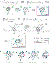
(a) Synthetic steps of nanoprobes Den-RGD and Den-RGD-Angio. (b) Schematics of the targeted and control nanoprobes. (Reprinted with permission from Ref . Copyright 2011 American Chemical Society)
FIGURE 7 |
FIGURE 7 |
Den-RGD-Angio demonstrated high BBB permeability and T/N ratio in vivo. (a) Representative T1-weighted magnetic resonance (MR) images of normal mouse brain (upper panel, arrow points to the cortex) and tumor-bearing brain (lower panel, arrows point to the tumor) before and at selected time PI of Den-RGD-Angio (0.05 mmol/kg [Gd3+], iv). Histological H&E staining verified the tumor boundary in MRI (bar, 2.0 mm). (b) In vivo time-dependent MR signal associated T/N ratio before and PI of nanoprobe (n = 4). Points present mean values and bars show the maximum and minimum values (data range). (c) Representative near-infrared fluorescence (NIRF) and X-ray/color-coded NIRF images of the normal mouse (left panel) and brain tumor-bearing mouse (right panel) at 24 h PI of Den-RGD-Angio (5.0 nmol/mouse based on dendrimer). (d) In vivo time-dependent NIR fluorescence T/N ratio (n = 3). Points present mean values and bars show the data range. (Reprinted with permission from Ref . Copyright 2011 American Chemical Society)

Similar articles

Cited by

References

    1. Brandes AA, Tosoni A, Franceschi E, Reni M, Gatta G, Vecht C. Glioblastoma in adults. Crit Rev Oncol Hematol 2008, 67:139 – 152. - PubMed
    1. Sanai N, Berger MS. Glioma extent of resection and its impact on patient out-come. Neurosurgery 2008, 62:753 – 764. - PubMed
    1. Stupp R, Mason WP, van den Bent MJ, Weller M, Fisher B, Taphoorn MJ, Belanger K, Brandes AA, Marosi C, Bogdahn U, et al. Radiotherapy plus concomitant and adjuvant temozolomide for glioblastoma. N Engl J Med 2005, 352:987 – 996. - PubMed
    1. Bucci MK, Maity A, Janss AJ, Belasco JB, Fisher MJ, Tochner ZA, Rorke L, Sutton LN, Phillips PC, Shu HK. Near complete surgical resection predicts a favorable outcome in pediatric patients with nonbrainstem, malignant gliomas: results from a single center in the magnetic resonance imaging era. Cancer 2004, 101:817 – 824. - PubMed
    1. Stupp R, Hegi ME, van den Bent MJ, Mason WP, Weller M, Mirimanoff RO, Cairncross JG. European Organisation for Research and Treatment of Cancer Brain Tumor and Radiotherapy Groups; National Cancer Institute of Canada Clinical Trials Group. Changing paradigms-an update on the multidisciplinary management of malignant glioma. Oncologist 2006, 11:165 – 180. - PubMed

Publication types

LinkOut - more resources