. 2013 Mar;70(3):134-47.
doi: 10.1002/cm.21095. Epub 2013 Feb 1.

Nonmyofilament-associated troponin T3 nuclear and nucleolar localization sequence and leucine zipper domain mediate muscle cell apoptosis

Affiliations

Nonmyofilament-associated troponin T3 nuclear and nucleolar localization sequence and leucine zipper domain mediate muscle cell apoptosis

Tan Zhang et al. Cytoskeleton (Hoboken). 2013 Mar.

Abstract

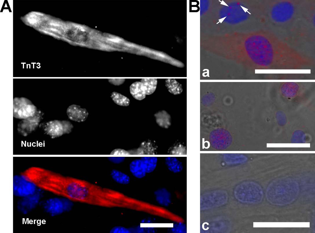
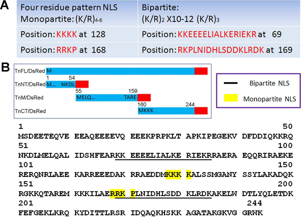
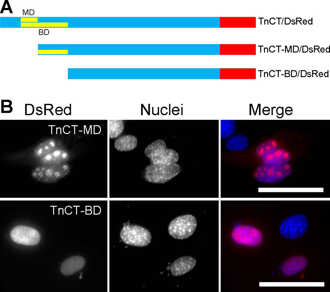
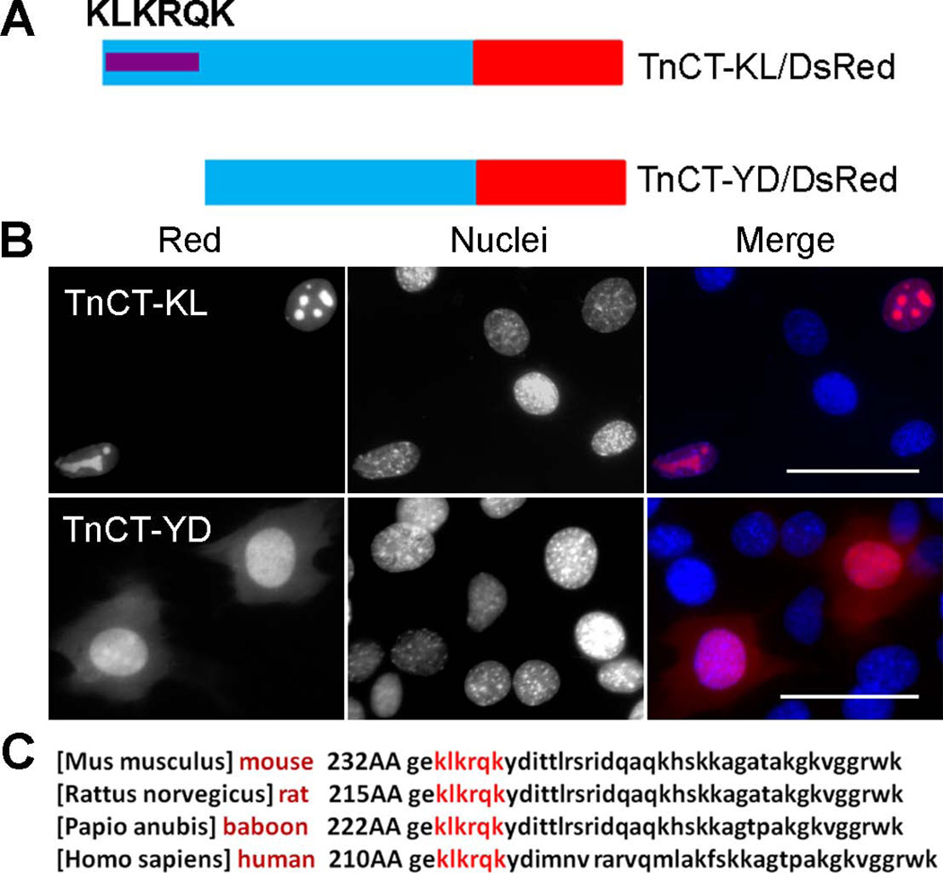
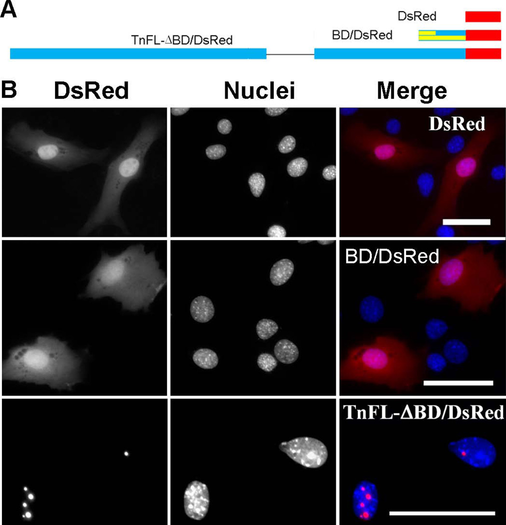
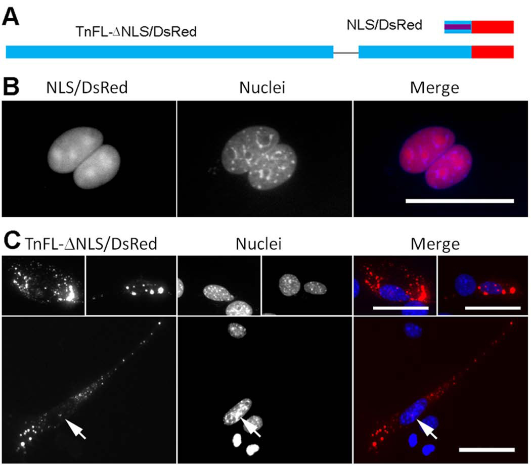
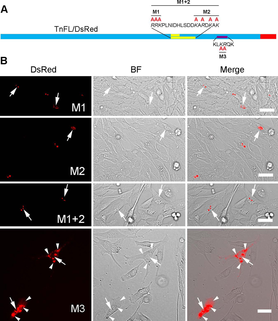
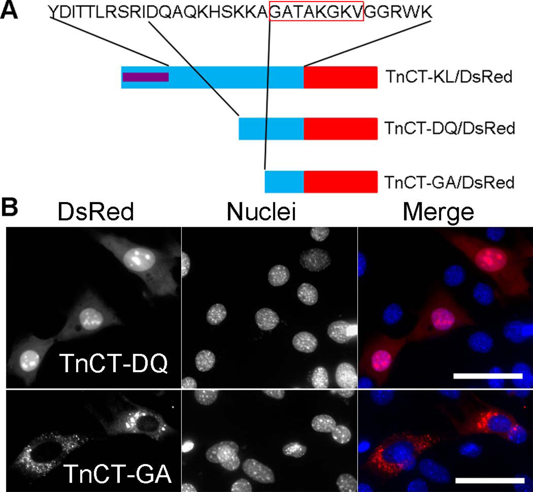
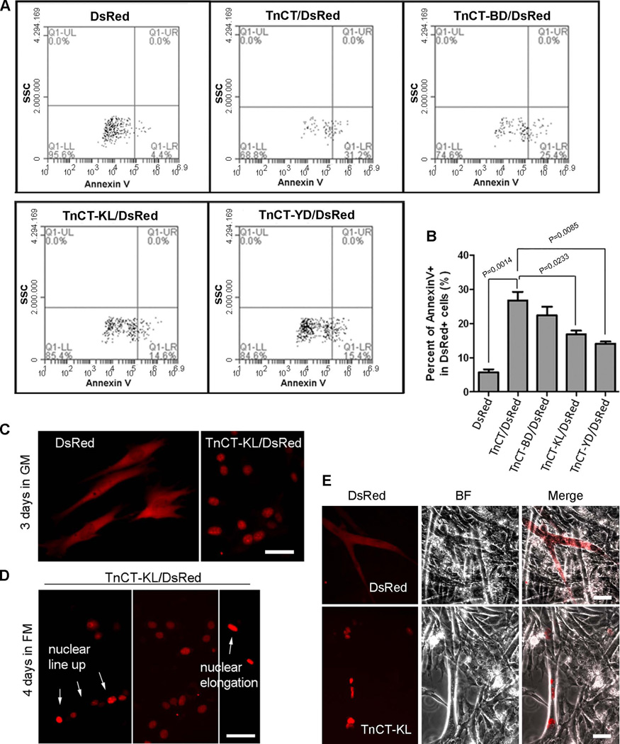
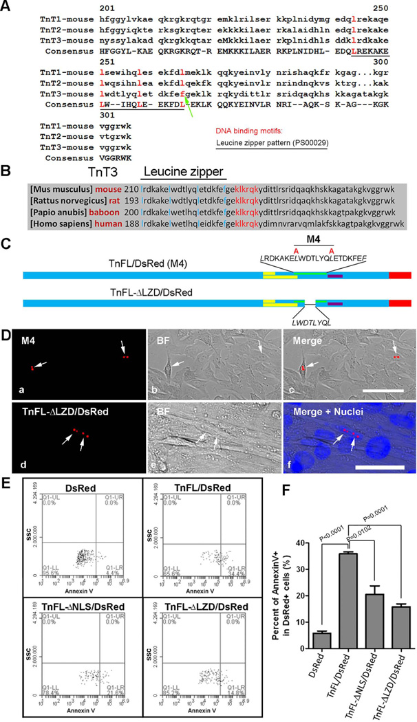
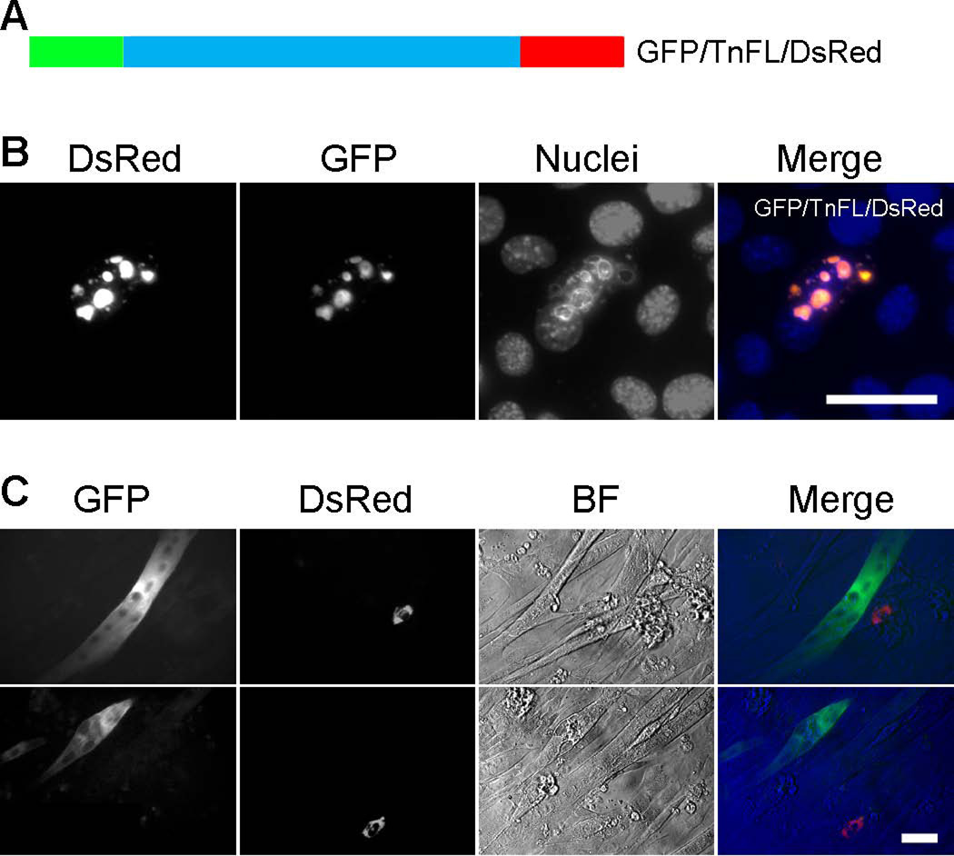
Troponin T (TnT) plays a major role in striated muscle contraction. We recently demonstrated that the fast skeletal muscle TnT3 isoform is localized in the muscle nucleus, and either its full-length or COOH-terminus leads to muscle cell apoptosis. Here, we further explored the mechanism by which it enters the nucleus and promotes cytotoxicity. Amino acid truncation and substitution showed that its COOH-terminus contains a dominant nuclear/nucleolar localization sequence (KLKRQK) and the basic lysine and arginine residues might play an important role in the nuclear retention and nucleolar enrichment of KLKRQK-DsRed fusion proteins. Deleting this domain or substituting lysine and arginine residues (KLAAQK) resulted in a dramatic loss of TnT3 nuclear and nucleolar localization. In contrast, the GATAKGKVGGRWK domain-DsRed construct localized exclusively in the cytoplasm, indicating that a nuclear exporting sequence is possibly localized in this region. Additionally, we identified a classical DNA-binding leucine zipper domain (LZD) which is conserved among TnT isoforms and species. Deletion of LZD or KLKRQK sequence significantly reduced cell apoptosis compared to full-length TnT3. We conclude that TnT3 contains both a nuclear localization signal and a DNA-binding domain, which may mediate nuclear/nucleolar signaling and muscle cell apoptosis.


Figures

Figure 1
Figure 1. Endogenous nuclear TnT3 in cultured C2C12 early in differentiation
C2C12 cells were immunostained with TnT3 antibody following two procedures: (A) Fixed prior to permeabilization. An elongated mononucleated myocyte was positively stained with TnT3 in both cytoplasmic and nuclear areas. Neighboring cells were not stained. (B) Permeabilized prior to fixation to allow extraction of cytoplasmic components. TnT3 was detected in some nuclear areas as isolated foci (a, arrows) or abundant punctates (a and b); a control, using only the secondary antibody, shows no positive staining (c). Nuclei (blue, Hoechst 33342) and TnT3 (red), and brightfield images are merged. Scale bars, 50 µm.
Figure 2
Figure 2. Prediction of TnT3 nuclear localization signal (NLS)
(A) Predicted monopartite and bipartite NLS sequence and positions. (B) Predicted NLS distribution along TnT3. Monopartite NLS is highlighted in yellow, and bipartite is underlined; one of each is localized in the TnM/DsRed and TnCT/DsRed constructs. The analysis used PSORT II.
Figure 3
Figure 3. Removal of the predicted monopartite and bipartite NLSs from TnCT/DsRed does not prevent its nuclear localization
(A) MD or BD-deleted TnT3 COOH-terminus fused to DsRed. (B) C2C12 cells expressing MD or BD deletion constructs, which both localized exclusively in the nucleus, but enrichment in the nucleolar area was reduced, particularly for TnCT-BD/DsRed. Nuclei (blue, Hoechst 33342), TnT3-DsRed fusion protein (red). Scale bar, 50 µm.
Figure 4
Figure 4. KLKRQK is needed for TnCT nuclear localization and is conserved among species
(A) Schematic representation of DsRed-fused TnT3 COOH-terminus mutant with the KLKRQK sequence retained (TnCT-KL/DsRed) or removed (TnCT-YD/DsRed). (B) C2C12 cells expressing the deletion constructs. TnCT-KL/DsRed is localized exclusively in the nucleus with an enriched nucleolar area. In contrast, TnCT-YD/DsRed is present throughout the cell, with marked reduction in nucleolar enrichment. Nuclei (blue, Hoechst 33342), TnT3-DsRed fusion protein (red). Scale bar, 50 µm. (C) The Tnnt3 KLKRQK sequence is conserved across mammalian species.
Figure 5
Figure 5. The predicted bipartite NLS does not play a major role in TnT3 nuclear localization
(A) Schematic representation of a DsRed-fused bipartite NLS alone (BD/DsRed) and bipartite NLS-deleted full-length TnT3 (TnFL-ΔBD/DsRed). (B) DsRed alone is localized throughout the C2C12 cell with no signs of nucleolar enrichment. BD/DsRed shows a subcellular localization similar to that of DsRed alone, with weak nucleolar enrichment. TnFL-ΔBD/DsRed shows an exclusive nucleolar enrichment like that described for wild-type TnFL/DsRed (Zhang et al. 2011). Nuclei (blue, Hoechst 33342), TnT3-DsRed fusion protein (red). Scale bar, 50 µm.
Figure 6
Figure 6. KLKRQK is a genuine NLS and plays a major role in TnT3 nuclear localization
(A) Schematic representation of DsRed-fused KLKRQK alone (NLS/DsRed) and full-length TnT3 devoid of this sequence (TnFL-ΔNLS/DsRed). (B) NLS/DsRed shows an exclusive nuclear localization with an enriched nucleolar area in C2C12 cells. (C) TnFL-ΔNLS/DsRed shows exclusion from the C2C12 nucleus, and only a minor fraction is localized in the nucleolar area (arrows). Nuclei (blue, Hoechst 33342), TnT3-DsRed fusion protein (red). Scale bar, 50 µm.
Figure 7
Figure 7. Basic residues in the KLKRQK sequence play a critical role in TnT3 nuclear localization
(A) Schematic representation of DsRed, full-length TnT3 fusion protein with K or R amino acid substitution by A in the MD, BD, and NLS regions. (B) Only mutating the NLS region greatly reduced its nuclear localization in C2C12 cells (M3). Arrows indicate the nucleus while arrowheads the cytoplasm. BF, brightfield. Scale bar, 50 µm.
Figure 8
Figure 8. NESs and NLSs in the TnT3 COOH-terminus
(A) Schematic representation of DsRed-fused TnT3 COOH-terminus deletion constructs. (B) TnCT-DQ/DsRed is localized throughout the C2C12 cell with an enriched nucleolar area, indicating a NLS/NoLS. In contrast, TnCT-GA/DsRed shows a dramatic exclusion from the nucleus, indicating a possible NES in the GATAKGKVGGRWK region, where the sequence in the red box overlaps with the predicted NES (Figure S1). Nuclei (blue, Hoechst 33342), TnT3-DsRed fusion protein (red). Scale bar, 50 µm.
Figure 9
Figure 9. TnT3 cytotoxicity is gradually reduced by truncating the TnT3 COOH-terminus
C2C12 cells transfected with various TnCT/DsRed constructs or DsRed alone for 48 hours were analyzed by flow cytometry. Data were collected on at least 50,000 freshly stained cells and at least 1000 DsRed positive cells and analyzed for staining with the cell death marker Annexin V (n=3). (A) Representative analyses of Annexin V staining after DsRed pregating. SSC, side scatter. (B) The percent of total apoptotic and necrotic cells (Annexin V+) was compared among different constructs. (C) A representative fluorescence image showing that C2C12 cells expressing TnCT-KL/DsRed or DsRed remain healthy in extended GM cultures and survive and fuse into myotubes in differentiation medium. Arrows indicate nuclear line up or elongation (D). (E) Brightfield merged with DsRed image showing that TnCT-KL/DsRed is localized in myotube nuclei, while DsRed alone is expressed throughout the myotube. Scale bars, 50 µm.
Figure 10
Figure 10. TnT3 Leucine Zipper Domain (LZD) is related to C2C12 cytotoxicity but not required for nuclear/nucleolar localization
(A) In silico identification of an LZD in TnT1 and TnT2 and sequence alignment showing that F replaces the 4th L in TnT3 (arrow). (B) Alignment of TnT3 COOH-terminal sequences showing that LZD is conserved across mammalian species. (C) Schematic representation of LZD partially deleted from full-length TnT3 (TnFL-DLZD/DsRed) and constructs with leucine mutated into alanine (M4). (D) LZD mutation (a-c) or truncation (d-f) does not affect nuclear localization and nucleolar enrichment in C2C12 myoblasts and myotubes. Nuclei (blue, Hoechst 33342), TnT3-DsRed fusion protein (red), brightfield (BF). Scale bars, 50 µm. (E) Representative analyses of Annexin V staining after DsRed pre-gating. C2C12 cells transfected with various TnFL/DsRed constructs or DsRed alone for 48 hours were analyzed by flow cytometry. Data were collected on at least 50,000 freshly stained cells and at least 1000 DsRed positive cells and analyzed for staining with the cell death marker Annexin V (n=3). SSC, side scatter. (F) Percent of Annexin V+ in C2C12 cells expressing DsRed, TnFL/DsRed, TnFL-DNLS/DsRed, and TnFL-DLZD/DsRed.
Figure 11
Figure 11. Fragmented GFP/TnFL/DsRed showed decreased cytotoxicity
(A) Schematic representation of GFP (NH2-terminal) and DsRed (COOH-terminal) full-length TnT3 double fluorescence construct (GFP/TnFL/DsRed). C2C12 cells cultured in GM for 2 days showed nucleolar GFP and DsRed fluorescence overlap, indicating nucleolar localization of intact TnT3 (B). No double fluorescence-positive cells survived culture in differentiation medium yet most showed either GFP (in myoblasts or myotubes) or DsRed (in myoblasts) fluorescence mainly in the cytoplasm (C). Scale bars, 50 µm.

Similar articles

Cited by

References

    1. Asumda FZ, Chase PB. Nuclear cardiac troponin and tropomyosin are expressed early in cardiac differentiation of rat mesenchymal stem cells. Differentiation. 2012;83(3):106–115. - PubMed
    1. Banfic H, Visnjic D, Mise N, Balakrishnan S, Deplano S, Korchev YE, Domin J. Epidermal growth factor stimulates translocation of the class II phosphoinositide 3-kinase PI3K-C2beta to the nucleus. Biochem J. 2009;422(1):53–60. - PubMed
    1. Bergmann O, Bhardwaj RD, Bernard S, Zdunek S, Barnabe-Heider F, Walsh S, Zupicich J, Alkass K, Buchholz BA, Druid H, et al. Evidence for cardiomyocyte renewal in humans. Science. 2009;324(5923):98–102. - PMC - PubMed
    1. Birbrair A, Wang ZM, Messi ML, Enikolopov GN, Delbono O. Nestin-GFP transgene reveals neural precursor cells in adult skeletal muscle. PLoS One. 2011;6(2):e16816. - PMC - PubMed
    1. Birbrair A, Zhang T, Wang ZM, Messi ML, Enikolopov GN, Mintz A, Delbono O. Skeletal muscle neural progenitor cells exhibit properties of NG2-glia. Exp Cell Res. 2012a - PMC - PubMed

Publication types

LinkOut - more resources