. 2013 Jun;29(5):1069-76.
doi: 10.1007/s10554-013-0187-9. Epub 2013 Feb 3.

Effects of respiratory motion on coronary wall MR imaging: a quantitative study of older adults

Affiliations

Effects of respiratory motion on coronary wall MR imaging: a quantitative study of older adults

Kai Lin et al. Int J Cardiovasc Imaging. 2013 Jun.

Abstract

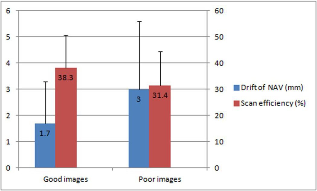
The aim of the present study is to assess the effects of respiratory motion on the image quality of two-dimensional (2D), free-breathing, black-blood coronary wall magnetic resonance (MR) imaging. This study was compliance with the HIPPA. With the approval of the institution review board, 230 asymptomatic participants, including 164 male patients (72.9 ± 4.4 years) and 66 female patients (72.4 ± 5.1 years), were recruited. Written informed consent was obtained. A 2D navigator (NAV)-gated, black-blood coronary wall MR imaging sequence was run on the left main artery, the left anterior descending artery and the right coronary artery. The drift of the location of the NAV and scan efficiency were compared between good (scored 2 or 3) and poor images (scored 1). Age, body weight, body weight index, heart rate, length of the rest period of cardiac motion, diaphragm excursion and breathing frequency were compared using a t test between the "successful" (having 2 or 3 good images) and "unsuccessful" cases (having 1 or 0 good images). A logistic regression model was applied to identify the contributors to good image quality. The drift of the NAV location and the scan efficiency were higher in the 411 good images compared with the 279 poor images. Minimal drift of the NAV location and low body weight were identified as independent predictors of good images after using a logistic regression model to adjust for multiple physiological and technical factors. The stability of respiratory motion significantly influences the image quality of 2D, free-breathing, black-blood coronary wall MR imaging.


Figures

Figure 1
Figure 1
Minimal drift of the NAV location and high scan efficiency were affiliated with good images.
Figure 2
Figure 2
A 76-year-old female with a 5-year history of HTN. Her heart rate was 58 beats/minute. The rest period of cardiac motion was 220 msec. Her body weight was 51.2 kg, and her BMI was 22.5. Her breathing frequency was 18 breaths/minute. The excursion of the diaphragm was 16 mm. a. For the LM, the initial location of the NAV was 127 mm. The green box is the acceptance window of the NAV (± 2.5 mm) and the red box is the search window for the diaphragm (± 32 mm). * b. The location of the NAV was 127 mm at the end of the scan. The drift of the NAV was 0 mm during the scan. The scan efficiency was 49%. c. The image quality of the LM was ranked as 3.
Figure 3
Figure 3
A 69-year-old male with an 8-year history of DM. His heart rate was 62 beats/minute. The rest period of cardiac motion was 200 msec. His body weight was 68.9 kg, and his BMI was 24.4. His breathing frequency was 15 breaths/minute. The excursion of the diaphragm was 21 mm. a. For the LM, the initial location of the NAV was 132 mm. b. The location of the NAV was 133 mm at the end of the scan. The drift of the NAV was 1 mm during the scan. The scan efficiency was 29%. c. The image quality of the LM was ranked as 2.
Figure 4
Figure 4
A 72-year-old, healthy female. Her heart rate was 52 beats/minute. The rest period of cardiac motion was 280 msec. Her body weight was 57.4 kg, and her BMI was 23.0. Her breathing frequency was 18 breaths/minute. The excursion of the diaphragm was 18 mm. a. For the LM, the initial location of the NAV was 123 mm. b. The location of the NAV was 120 mm at the end of the scan. The drift of the NAV was 3 mm during the scan. The scan efficiency was 24%. c. The image quality of the LM was ranked as 1. * The presentation of the location of the NAV on MR scanners may be vendor-specified.

References

    1. Roger VL, Go AS, Lloyd-Jones DM, et al. Heart Disease and Stroke Statistics--2012 Update: A Report From the American Heart Association. Circulation. 2012;125:e2–e220. - PMC - PubMed
    1. Nicholls SJ, Ballantyne CM, Barter PJ, et al. Effect of two intensive statin regimens on progression of coronary disease. N Engl J Med. 2011;365:2078–2087. - PubMed
    1. Fayad ZA, Mani V, Fuster V. The time has come for clinical cardiovascular trials with plaque characterization as an endpoint. Eur Heart J. 2012;33:160–161. - PMC - PubMed
    1. Fayad ZA, Fuster V, Fallon JT, et al. Noninvasive in vivo human coronary artery lumen and wall imaging using black-blood magnetic resonance imaging. Circulation. 2000;102:506–510. - PubMed
    1. Miao C, Chen S, Macedo R, et al. Positive remodeling of the coronary arteries detected by magnetic resonance imaging in an asymptomatic population: MESA (Multi-Ethnic Study of Atherosclerosis) J Am Coll Cardiol. 2009;53:1708–1715. - PMC - PubMed

Publication types

LinkOut - more resources