. 2013 Mar 15;73(6):1981-92.
doi: 10.1158/0008-5472.CAN-12-2962. Epub 2013 Feb 1.

FOXC2 expression links epithelial-mesenchymal transition and stem cell properties in breast cancer

Affiliations

FOXC2 expression links epithelial-mesenchymal transition and stem cell properties in breast cancer

Brett G Hollier et al. Cancer Res. .

Abstract

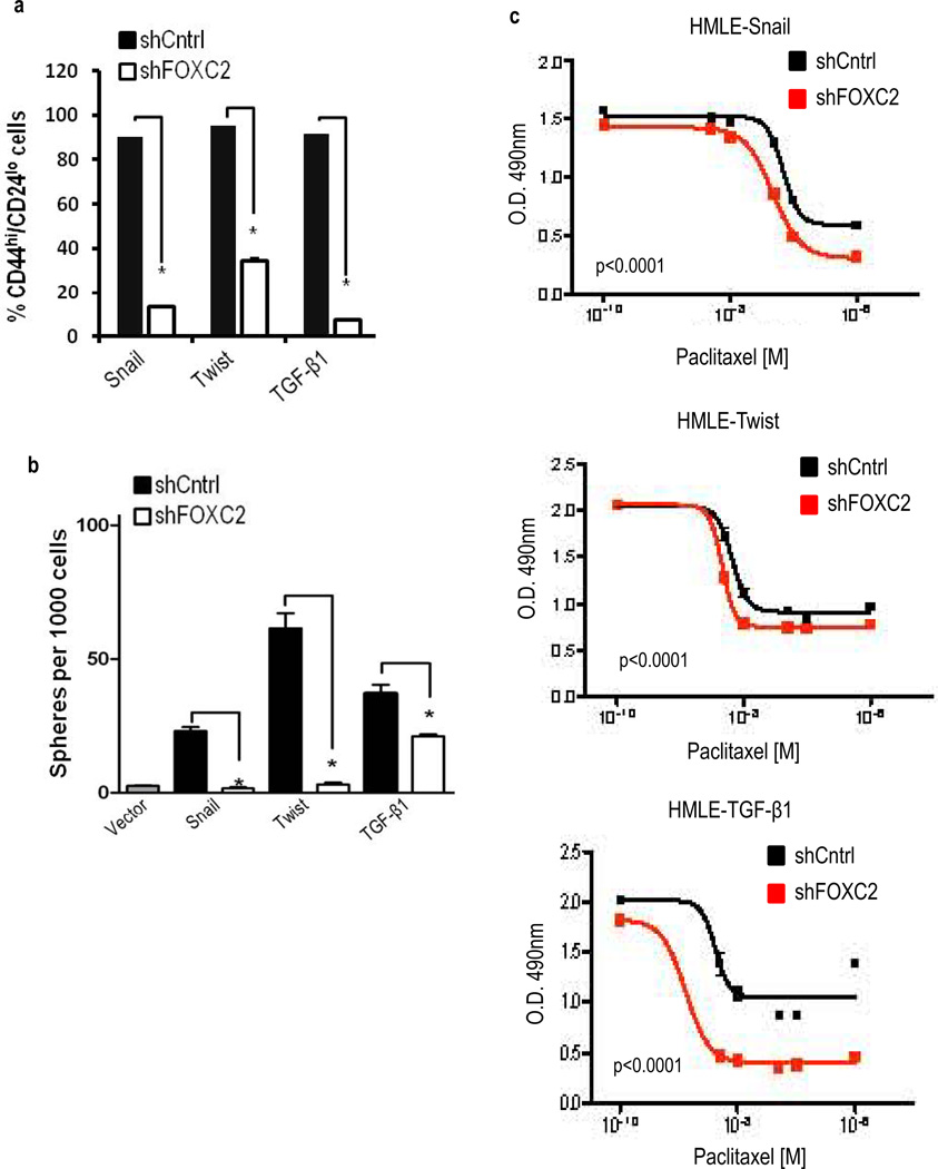
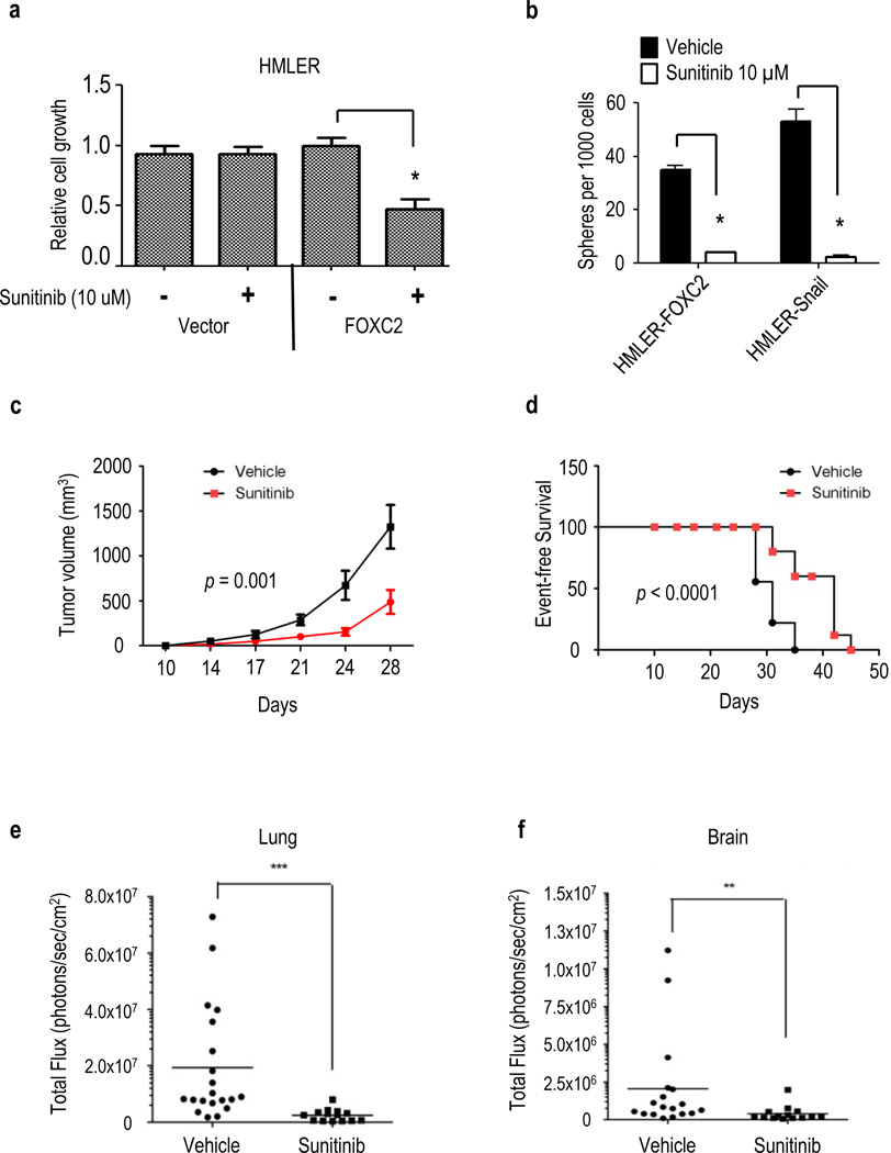
Resistance to chemotherapy and metastases are the major causes of breast cancer-related mortality. Moreover, cancer stem cells (CSC) play critical roles in cancer progression and treatment resistance. Previously, it was found that CSC-like cells can be generated by aberrant activation of epithelial-mesenchymal transition (EMT), thereby making anti-EMT strategies a novel therapeutic option for treatment of aggressive breast cancers. Here, we report that the transcription factor FOXC2 induced in response to multiple EMT signaling pathways as well as elevated in stem cell-enriched factions is a critical determinant of mesenchymal and stem cell properties, in cells induced to undergo EMT- and CSC-enriched breast cancer cell lines. More specifically, attenuation of FOXC2 expression using lentiviral short hairpin RNA led to inhibition of the mesenchymal phenotype and associated invasive and stem cell properties, which included reduced mammosphere-forming ability and tumor initiation. Whereas, overexpression of FOXC2 was sufficient to induce CSC properties and spontaneous metastasis in transformed human mammary epithelial cells. Furthermore, a FOXC2-induced gene expression signature was enriched in the claudin-low/basal B breast tumor subtype that contains EMT and CSC features. Having identified PDGFR-β to be regulated by FOXC2, we show that the U.S. Food and Drug Administration-approved PDGFR inhibitor, sunitinib, targets FOXC2-expressing tumor cells leading to reduced CSC and metastatic properties. Thus, FOXC2 or its associated gene expression program may provide an effective target for anti-EMT-based therapies for the treatment of claudin-low/basal B breast tumors or other EMT-/CSC-enriched tumors.


Figures

Figure 1
Figure 1
FOXC2 expression is necessary to maintain the mesenchymal and invasive properties induced by EMT in mammary epithelial cells. (a) Phase-contrast images of HMLE-Vector, - Snail, - Twist and -TGF-β1 cells expressing either control-shRNA (shCntrl) or FOXC2-shRNA (shFOXC2). Scale bar indicates 100 µm. (b) Expression of EMT marker mRNA by quantitative RT-PCR. GAPDH was used as an internal control. n=3; error bars indicate SEM. N.D. = not detected. (c) Western blot analysis of EMT marker protein expression upon FOXC2 suppression in HMLE-Snail, -Twist and -TGF-β1 cells, respectively. (d) Quantification of invasion in Matrigel Transwell chambers using HMLE-Snail-shCntrl and HMLE-Snail-shFOXC2 cells in response to basic fibroblast growth factor (bFGF) and platelet-derived growth factor-BB (PDGF-BB). n=3; error bars indicate SEM. * P<0.05. (e) Confocal microscopy images of HMLE-Snail-shCntrl and HMLE-Snail-shFOXC2 cells in 3D lrECM cultures. The integrity of the basement membrane was assessed using anti-Laminin V (red) with DAPI nuclear stain (blue). Scale bar indicates 100 µm.
Figure 2
Figure 2
FOXC2 expression is required for EMT-derived stem cell properties in mammary epithelial cells. (a) Quantification of CD44 (CD44-PE) and CD24 (CD24-FITC) expression by FACS analysis in HMLE-Snail, -Twist and -TGF-β1 cells expressing either shCntrl or shFOXC2. n=3; error bars indicated SEM. * P<0.05. (b). In vitro quantification of mammospheres formed by cells described in (a). n=3, error bars indicate SEM. * P<0.05. (c) Quantification of cell viability using an MTS assay in cells described in (a) following culture for 96 hours in increasing concentrations of Paclitaxel. Data is represented as the absorbance (O.D) at 490 nm. n=3, p < 0.005.
Figure 3
Figure 3
FOXC2 expression is increased in stem cell enriched populations and is sufficient to promote phenotypes associated with CSCs. (a) Western blot analysis of FOXC2 expression in the stem cell enriched CD44hi/CD24lo (44hi/24lo) and more differentiated CD44lo/CD24hi (44lo/24hi) cellular fractions isolated by FACS from HMLER and SUM159 cell lines. (b) Western blot analysis of FOXC2 expression in cells cultured in monolayer culture (2D) and stem cell enriched mammosphere cultures (MS) for the indicated breast cancer cell lines. (c) FACS analysis of CD44 and CD24 expression in HMLER-Vector and HMLER-FOXC2 cells. Representative FACS plots are shown. (d) In vitro quantification of mammospheres formed by 1000 cells described in (c) (n = 3); error bars indicate SEM. * P<0.05. (e) Quantification of cell viability by MTS assay using HMLER cells expressing either Vector or FOXC2 cDNA following culture for 96 hours in increasing concentrations of Paclitaxel. Data represented is the mean absorbance (O.D) at 490 nm, (n = 3). (f) Tumor incidence of FOXC2 expressing HMLER cells injected into the mammary fat pad of NOD/SCID mice in limiting dilutions. (g) Tumor growth quantification of luciferase labelled HMLER-Vector and HMLER-FOXC2 xenografts in vivo using bioluminescence after 28 days of inoculation into the mammary fat pad of NOD/SCID mice. (n = 5). (h) Ex vivo bioluminescence images of the indicated organs of mice carrying HMLER-Vector and HMLER-FOXC2 xenografts after 28 days.
Figure 4
Figure 4
FOXC2 derived gene signature is enriched in claudin-low human breast cancer samples and can accurately predict claudin-low human tumors. (a,b) Measurement of FOXC2 gene expression signature (GES) in: (a) MDA-MB231 (GSE12237) and (b) CN34 (GSE12237) xenograft models consisting of the parental tumors and brain metastases. The box plots show the mean, 5%, and 95% distribution of the level of FOXC2 signature (42) in GSE12237. Predicted activation of FOXC2 between the primary tumors and metastases was compared and a p-value was calculated using a Student’s t-test. (c,d) The FOXC2 GES was scored in (c) tumors (GSE18229) and (d) established breast cancer cell lines (E-TABM-157). The box plots represent the mean, 5%, and 95% distribution of the FOXC2 signature scores across the (c) breast tumor subtypes data derived from GSE18229 (35) and (d) an expression dataset of 51 breast cancer cell lines described in (43) (E-TABM-157; ArrayExpress). The one-way ANOVA significance for each plot was p < 0.0001. (e) Western blot analysis of FOXC2 expression in a panel of established breast cancer cell lines representing luminal, basal and claudin-low subtypes.
Figure 5
Figure 5
Attenuation of FOXC2 expression reduces the mesenchymal and stem cell properties of breast cancer cell lines with a claudin-low phenotype. (a) Phase-contrast images of SUM159 and HMLER-Snail cells expressing either a control-shRNA (shCntrl) or FOXC2-shRNA (shFOXC2). Scale bar indicates 100 µm. (b) Western blot analysis of EMT marker protein expression upon FOXC2 suppression in SUM159 and HMLER-Snail cells. (c,d) Quantification of Transwell cell migration for SUM159 (c) and HMLER-Snail (d) cells expressing either a control-shRNA (shCntrl) or FOXC2-shRNA (shFOXC2) in response to epidermal growth factor (EGF). Columns represent the average cell migration (n = 6) relative to that induced in shCntrl cells by serum free media alone (SFM). Error bars indicate SEM. * P<0.05. (e) Confocal microscopy images of SUM159-shCntrl and SUM159-shFOXC2 cells in 3D lrECM cultures. Cell invasion was qualitatively assessed using anti-vimentin (green) and F-actin detected with TRITC-conjugated phalloidin (red). Nuclei were stained with DAPI (blue). Scale bar indicates 250 µm. (f) Quantification of FACS analysis of CD44 (CD44-PE) and CD24 (CD24-FITC) expression in SUM159, MDA-MB-231 and HMLER-Snail cells expressing either shCntrl or shFOXC2, (n = 3) error bars indicated SEM. * P<0.05. (g) In vitro quantification of mammospheres formed by 1000 cells described in (f), (n = 3), error bars indicate SEM. * P<0.05. (h) Tumor incidence of SUM159 and HMLER-Snail cells expressing shCntrl or shFOXC2 injected into the mammary fat pad of NOD/SCID mice in limiting dilutions and measured as palpable tumors after 12 weeks.
Figure 6
Figure 6
FOXC2 regulates the expression of PDGFR-β. (a) Western blot analysis of PDGFR-β expression in normal (i) and transformed (ii) mammary epithelial cells following EMT induction by multiple factors as well as in stem cell enriched (44hi/24lo) relative to (44lo/24hi) (iii) and mammosphere cultures (iv). (b) Western blot analysis of PDGFR-β expression in a panel of established breast cancer cell lines representing luminal, basal and claudin-low subtypes. (c) Quantification of Transwell cell migration for the indicated cell lines in response to the PDGFR-β ligand, platelet-derived growth factor-BB (PDGF-BB) (20 ng/ml), (n = 6) error bars indicate SEM, * P<0.05. (d) Phase contrast images of HMLER-Vector and HMLER-FOXC2 cells in 3D lrECM cultures in the presence and absence of PDGF-BB. Scale bar indicates 50 µm. (e,f) Western blot analysis of PDGFR-β expression in cells induced to undergo EMT by ectopic expression of Twist, Snail or TGF-β1 in HMLE derived cells (e) as well as in transformed cell lines (SUM159 and HMLER-Snail) (f) expressing either shCntrl or shFOXC2. (g) Quantification of Transwell cell migration for the indicated cell lines expressing either a control-shRNA (shCntrl) or FOXC2-shRNA (shFOXC2) in response to PDGF-BB (20 ng/ml), (n = 6) error bars indicate SEM, * P<0.05. (h) Quantification of binding of FOXC2 to the PDGFR-β promoter by ChIP assays, (n = 3) error bars represent the SEM. Capitalized nucleotides indicate the predicted FOXC2 binding sites at the indicated chromosomal locations.
Figure 7
Figure 7
Sunitinib inhibits the growth and metastasis of FOXC2-expressing tumors. (a) Quantification of HMLER-Vector and HMLER-FOXC2 cell viability in the presence of sunitinib (10 µM) relative to vehicle (DMSO), (n = 3), error bars indicate SEM, * P<0.05. (b) In vitro quantification of mammospheres formed by 1000 HMLER-FOXC2 and HMLER-Snail cells in the presence of sunitinib (10 µM) or DMSO, (n = 3), error bars indicate SEM, * P<0.05. (c) Tumor volume of HMLER-FOXC2 cells injected into the mammary fat pad of NOD/SCID mice and treated with 40 mg/kg of sunitinib or vehicle (n = 9) daily for the indicated number of days. (d) Event-free survival of mice with orthotopic HMLER-FOXC2 xenografts treated daily with sunitinib (40 mg/kg, n = 7) or vehicle (n = 10). Mice were euthanized once tumors reached 1.5 cm3. (e,f) Thirty days following initiation of sunitinib or vehicle treatment, mice were euthanized and the organs, (e) lung and (f) brain, were dissected and analysed for metastatic tumor burden using bioluminescence imaging. The luminescent signal of tumor cells is represented as the total photon flux detected in each organ from individual mice with the bar indicating the average. ***P < 0.001, **P< 0.05 compared to the vehicle control group.

References

    1. Creighton CJ, Li X, Landis M, et al. Residual breast cancers after conventional therapy display mesenchymal as well as tumor-initiating features. Proc Natl Acad Sci U S A. 2009;106:13820–13825. - PMC - PubMed
    1. Abraham BK, Fritz P, McClellan M, Hauptvogel P, Athelogou M, Brauch H. Prevalence of CD44+/CD24−/low cells in breast cancer may not be associated with clinical outcome but may favor distant metastasis. Clin Cancer Res. 2005;11:1154–1159. - PubMed
    1. Al-Hajj M, Wicha MS, Benito-Hernandez A, Morrison SJ, Clarke MF. Prospective identification of tumorigenic breast cancer cells. Proc Natl Acad Sci U S A. 2003;100:3983–3988. - PMC - PubMed
    1. Ginestier C, Hur MH, Charafe-Jauffret E, et al. ALDH1 Is a Marker of Normal and Malignant Human Mammary Stem Cells and a Predictor of Poor Clinical Outcome. Cell Stem Cell. 2007;1:555–567. - PMC - PubMed
    1. Liu R, Wang X, Chen GY, et al. The prognostic role of a gene signature from tumorigenic breast-cancer cells. N Engl J Med. 2007;356:217–226. - PubMed

Publication types

Substances