. 2013 Apr;64(6):1553-64.
doi: 10.1093/jxb/ert020. Epub 2013 Feb 1.

Lamin-like analogues in plants: the characterization of NMCP1 in Allium cepa

Affiliations

Lamin-like analogues in plants: the characterization of NMCP1 in Allium cepa

Malgorzata Ciska et al. J Exp Bot. 2013 Apr.

Abstract

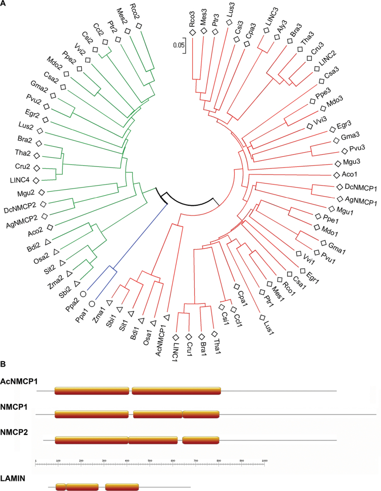
The nucleoskeleton of plants contains a peripheral lamina (also called plamina) and, even though lamins are absent in plants, their roles are still fulfilled in plant nuclei. One of the most intriguing topics in plant biology concerns the identity of lamin protein analogues in plants. Good candidates to play lamin functions in plants are the members of the NMCP (nuclear matrix constituent protein) family, which exhibit the typical tripartite structure of lamins. This paper describes a bioinformatics analysis and classification of the NMCP family based on phylogenetic relationships, sequence similarity and the distribution of conserved regions in 76 homologues. In addition, NMCP1 in the monocot Allium cepa characterized by its sequence and structure, biochemical properties, and subnuclear distribution and alterations in its expression throughout the root were identified. The results demonstrate that these proteins exhibit many similarities to lamins (structural organization, conserved regions, subnuclear distribution, and solubility) and that they may fulfil the functions of lamins in plants. These findings significantly advance understanding of the structural proteins of the plant lamina and nucleoskeleton and provide a basis for further investigation of the protein networks forming these structures.


Figures

Fig. 1.
Fig. 1.
Classification of NMCPs: evolutionary relationships and predicted protein structures. (A) Phylogenetic relationship of NMCPs inferred using the neighbour-joining method. Evolutionary distances were calculated using the p-distance method and are presented as the number of amino-acid differences per site. The phylogenetic tree is drawn to scale. The sequences classified as type 1 NMCP are marked in red and type 2 are in green, with the two members in Physcomitrella patens in blue. Dicotyledon species are represented by rhombi; monocotyledons by triangles; and moss by circles. Sequence accession data are shown in Supplementary Table S1. (B) Schematic representation of the coiled-coil prediction (MARCOIL) for AcNMCP1, typical NMCP1 and NMCP2, and lamin (orange boxes).
Fig. 2.
Fig. 2.
Conserved regions and phosphorylation sites in AcNMCP1, NMCP1, and NMCP2. (A) Schematic representation of conserved regions, predicted nuclear localization signals (green boxes) and phosphorylation sites (red bars, cdk1; grey bar, PKA/PKG). Localization of the conserved regions is indicated by green bars with corresponding numbers. Coiled coils are represented as orange boxes. (B) MEME motifs displayed as ‘sequence LOGOS’. The height of each letter reflects the probability of its localization at this position. Letters are coloured using the same colour scheme as the MEME motifs based on the biochemical properties of the amino acids.
Fig. 3.
Fig. 3.
Characterization of AcNMCP1. (A) Immunoblot detection of proteins using anti-AcNMCP1 in Zma (corn), Psa (pea), Ath (Arabidopsis thaliana), Tae (wheat), Sce (rye), Asa (garlic), and Ace (onion). Ace’, overexposure of Ace; (–), negative control with primary antibody omitted. (B) Detection of AcNMCP1 in onion nuclear fractions extracted in 2 × Laemmli buffer (SDS), 7M urea/2M thiourea (7M U), and 6M guanidine thiocyanate (GITC). (C, D) 2D-immunoblots of A. cepa whole-nuclear extracts (C) and total Arabidopsis protein (D), probed with the anti-AcNMCP1 antibody.
Fig. 4.
Fig. 4.
Subnuclear localization of AcNMCP1. (A–E) Confocal sections of meristematic nuclear fractions after incubation with the anti-AcNMCP1 antibody, demonstrating the distribution of the protein along the nuclear periphery (A–E) and in the nucleoplasm on occasion (D, E). (B’’) High magnification of a portion of the nucleus in B showing the punctuate-like distribution of the peripheral labelling. (C) Negative control incubated with the secondary antibody alone. (A’, B’, C’, D’, and E’) Overlay of the corresponding anti-NMCP1- and DAPI-stained images. (F) High-resolution pre-embedding immunogold labelling. Portion of a nucleus that exhibit accumulations of gold particles in the peripheral plant lamina (thick arrows) and scarce labelling in the interchromatin domains (id) (thin arrows). The condensed chromatin masses (chr) and nucleolus (No) showed no labelling. Bar in F = 100nm.
Fig. 5.
Fig. 5.
AcNMCP is a component of the nucleoskeleton (NSK). (A) Detection of AcNMCP1 in the nuclear (N), insoluble (F1, F2, NSK), and soluble (S1, S2, S3) fractions obtained during NSK extraction in immunoblots probed with anti-AcNMCP1. The 200-kDa band of AcNMCP1 was present in all the insoluble fractions but not in the soluble fractions. (B) Coomassie blue staining of a gel run in parallel showing the complex protein composition of the insoluble and soluble fractions. (C, D) Confocal images of NSKs showing the predominant accumulation of AcNMCP1 in the lamina and weaker staining associated with the internal NSK. (C’, D’) Differential interference contrast images of the corresponding fields. (E) Immunogold labelling of NSK showing the association of gold particles with the plant lamina and internal NSK. Bars, 25 µm (C, C’), 10 µm (D, D’), 100nm (E).
Fig. 6.
Fig. 6.
Expression and distribution of AcNMCP1 in nuclei isolated from different root cell types. (A) Localization of the onion root zones used in this analysis and their corresponding DNA content determined by flow cytometry. (B) AcNMCP1 levels detected by immunoblotting with the anti-AcNMCP1 antibody. AcNMCP1 expression was abundant in the proliferating (m) and quiescent (q) meristems, although it decreased significantly in non-meristematic cells (e = elongation zone; d = differentiated zone). H1 histones stained with Coomasie blue were used as loading controls. (C) Peripheral (qP, mP, dP) and central (qC, mC, dC) confocal sections showing the distribution of AcNMCP1 in the periphery and nuclear interior of quiescent (q) and proliferating (m) meristems and in differentiated cells (d). Arrows in qC point to the nucleoplasmic aggregates of the protein in quiescent meristems and arrows in dP to the gaps in the peripheral distribution of the protein in differentiated cells. qP’, qC’, mP’, mC’, dP’, and dC’ show overlays of AcNMCP1 and DAPI staining.

References

    1. Bailey TL, Boden M, Buske FA, Frith M, Grant CE, Clementi L, Ren J, Li WW, Noble WS. 2009. MEME SUITE: tools for motif discovery and searching. Nucleic Acids Research 37, W202–W208 - PMC - PubMed
    1. Berkelman T. 2008. Quantitation of protein in samples prepared for 2-D electrophoresis. Methods in Molecular Biology 424, 43–49 - PubMed
    1. Blumenthal SS, Clark GB, Roux SJ. 2004. Biochemical and immunological characterization of pea nuclear intermediate filament proteins. Planta 218, 965–975 - PubMed
    1. Broers JL, Machiels BM, Kuijpers HJ, Smedts F, van den Kieboom R, Raymond Y, Ramaekers FC. 1997. A- and B-type lamins are differentially expressed in normal human tissues. Histochemistry and Cell Biology 107, 505–517 - PubMed
    1. Dechat T, Adam SA, Taimen P, Shimi T, Goldman RD. 2010a. Nuclear lamins. Cold Spring Harbor Perspectives in Biology 2, a000547 - PMC - PubMed

Publication types

MeSH terms