. 2013 Mar 15;190(6):2756-66.
doi: 10.4049/jimmunol.1202697. Epub 2013 Feb 1.

B1a cells enhance susceptibility to infection with virulent Francisella tularensis via modulation of NK/NKT cell responses

Affiliations

B1a cells enhance susceptibility to infection with virulent Francisella tularensis via modulation of NK/NKT cell responses

Deborah D Crane et al. J Immunol. .

Abstract

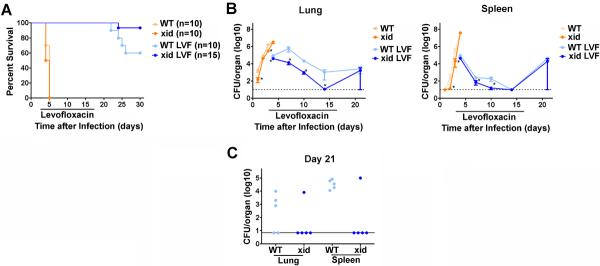
B1a cells are an important source of natural Abs, Abs directed against T-independent Ags, and are a primary source of IL-10. Bruton's tyrosine kinase (btk) is a cytoplasmic kinase that is essential for mediating signals from the BCR and is critical for development of B1a cells. Consequentially, animals lacking btk have few B1a cells, minimal Ab responses, and can preferentially generate Th1-type immune responses following infection. B1a cells have been shown to aid in protection against infection with attenuated Francisella tularensis, but their role in infection mediated by fully virulent F. tularensis is not known. Therefore, we used mice with defective btk (CBA/CaHN-Btk(XID)/J [XID mice]) to determine the contribution of B1a cells in defense against the virulent F. tularensis ssp. tularensis strain SchuS4. Surprisingly, XID mice displayed increased resistance to pulmonary infection with F. tularensis. Specifically, XID mice had enhanced clearance of bacteria from the lung and spleen and significantly greater survival of infection compared with wild-type controls. We revealed that resistance to infection in XID mice was associated with decreased numbers of IL-10-producing B1a cells and concomitant increased numbers of IL-12-producing macrophages and IFN-γ-producing NK/NKT cells. Adoptive transfer of wild-type B1a cells into XID mice reversed the control of bacterial replication. Similarly, depletion of NK/NKT cells also increased bacterial burdens in XID mice. Together, our data suggest B cell-NK/NKT cell cross-talk is a critical pivot controlling survival of infection with virulent F. tularensis.


Figures

Figure 1
Figure 1. XID mice exhibit enhanced survival and control of SchuS4 replication compared to WT mice
Mice were intranasally infected with 50 CFU F. tularensis strain SchuS4. As indicated, animals received 5 mg/kg levofloxacin (LVF) on days 3–16 of infection. CBA/J (WT) and CBA/CaHN-BtkXID/J (XID) mice were monitored for survival (A) or euthanized at the indicated time points for assessment of bacterial loads in the lung and spleen (B and C; n=4–5 mice/group/time point). (A) represents the results of two experiments pooled together. (B) is representative of two experiments of similar design. * = significantly less than WT mice (p<0.05). Error bars represent SEM.
Figure 2
Figure 2. XID mice have delayed and lower titers of antibodies directed against SchuS4
Mice (n= 4–5/group) were intranasally infected with 50 CFU F. tularensis strain SchuS4. As indicated, animals received 5 mg/kg levofloxacin on days 3–16 of infection. At the indicated time points CBA/J (WT) and CBA/CaHN-BtkXID/J (XID) mice were euthanized and serum collected. Serum was serially diluted and assessed for IgM (A) and IgG (B) directed against SchuS4 antigens by ELISA. Diluted serum was also assessed for its ability to agglutinate viable SchuS4 organisms (C). Error bars represent SEM. Data is representative of two experiments of similar design.
Figure 3
Figure 3. Macrophages from XID and WT mice are not different in their interaction with SchuS4
Macrophages were collected by bronchoalveolar lavage (alveolar) or differentiated from the bone marrow (Bone Marrow) and infected with SchuS4 at a MOI = 10. (A) At the indicated time points cells were lysed and intracellular bacteria were enumerated by plating lysates on MMH agar. (B) Twenty-four hours after infection cells were treated with Pam3CSK4. Supernatants were harvested 12 hours later and assessed for TNF-α by ELISA. Untreated, uninfected cells served as negative controls. Uninfected cells treated with Pam3CSK4 served as positive controls. *= significantly less than uninfected, Pam3CSk4 treated cells (p<0.05). Error bars represent SEM. Data is representative of three experiments similar design.
Figure 4
Figure 4. XID and WT mice do not have differences in recruitment of inflammatory cells following SchuS4 infection
Mice (n=4–5/group) were intranasally infected with 50 CFU F. tularensis strain SchuS4. As indicated, animals received 5 mg/kg levofloxacin on days 3–6 of infection. Animals were euthanized on day 7 of infection and assessed for the indicated cell populations by flow cytometry. Monocytes were characterized as CD11b+/Ly6C+/MHCII/Ly6G. Granulocytes were characterized as Ly6G+/MHCII. Error bars represent SEM. Data is representative of two experiments of similar design.
Figure 5
Figure 5. Control of SchuS4 infection in XID mice is correlated with fewer IL-10+ B1a cells and increased IL-12+ macrophages
Mice (n=4–5/group) were intranasally infected with 50 CFU F. tularensis strain SchuS4. As indicated, animals received 5 mg/kg levofloxacin on days 3–6 of infection. Animals were euthanized on day 7 of infection and single cell suspensions of the lung and spleen were generated. Cells were incubated with PMA, ionomycin and BFA for 4 hours were stained for specific surface receptors, permeabilized and stained for IL-10 and IL-12 and assessed for specific cell populations by flow cytometry. B1a cells were characterized as CD5Y/CD19Y. Macrophages were characterized as CD11b+/F480Y. Error bars represent SEM. * = significantly greater than uninfected XID (p<0.05). ** = significantly greater than uninfected WT and all XID (p<0.05). *** = significantly greater than all other groups (p<0.05). Data is representative of two experiments of similar design.
Figure 6
Figure 6. B1a cells derived IL-10 inhibits IL-12p40
Peritoneal exudate cells were collected from resting WT mice and were cultured with killed SchuS4 overnight. As indicated, some cells were either left untreated (−), or were incubated with rat immunoglobulin (Ig) or neutralizing anti-IL-10 antibodies (both at 10 ug/ml). Cultures were assessed for IL-10 and IL-12p40. * = significantly different untreated and Ig controls (p<0.05). Error bars represent SEM. Data is pooled from three experiments of similar design.
Figure 7
Figure 7. B1a cells contribute to exacerbation of SchuS4 infection
B1a cells were collected from the peritoneal cavities of CBA/J (wild type) mice and enriched by FACS. One day prior to infection XID mice (n=4–5/group) were injected with freshly isolated wild type B1a cells or PBS (untreated). Mice were intranasally infected with 50 CFU F. tularensis strain SchuS4. Animals received 5 mg/kg levofloxacin on days 3–6 of infection. (A) Mice were euthanized on day 7 of infection and bacteria were enumerated from the lung and spleen. (B) Survival of mice receiving B1a cells versus untreated controls. * = significantly different untreated controls (p<0.05). Error bars represent SEM. Data is representative of three experiments of similar design.
Figure 8
Figure 8. XID mice have significantly more IFN-γ+ NK/NKT cells after SchuS4 infection compared to WT mice
Mice (n=4–5/group) were intranasally infected with 50 CFU F. tularensis strain SchuS4. As indicated, animals received 5 mg/kg levofloxacin on days 3–6 of infection. Animals were euthanized on day 7 of infection and single cell suspensions of the lung and spleen were generated. Cells were incubated with PMA, ionomycin, and BFA for 4 hours, were stained for specific surface receptors, permeabilized and stained for IFN-γ and assessed for specific cell populations by flow cytometry. Error bars represent SEM. * = significantly greater than uninfected WT (p<0.05). ** = significantly greater than all other groups (p<0.05). Data is representative of two experiments of similar design.
Figure 9
Figure 9. NK/NKT cells contribute to control of SchuS4 infection in XID mice
Mice were treated with anti-asialo GM1 (anti-AGM1) antibodies to deplete NK/NKT cells or rabbit immunoglobulin (Ig) as a negative control on days −2, +1, +4, +7, +10. +13, +16, +19 of infection. Mice were intranasally infected with 50 CFU F. tularensis strain SchuS4. Animals received 5 mg/kg levofloxacin on days 3–6 of infection. All mice were euthanized on day 7 and 21 of infection and bacteria were enumerated from the lung and spleen. * = significantly different Ig treated controls (p<0.05). Error bars represent SEM. Data is representative of two experiments of similar design.
Figure 10
Figure 10. The role of B1a cells in tularemia
(A) Resident macrophages produce IL-12 in response to dead and/or lysing SchuS4 following antibiotic therapy. (B) IL-12 provokes NK/NKT cells to produce IFN-γ, which then (C) activates newly infected macrophages to kill SchuS4. However, B1a cells secrete IL-10 in response to dead bacteria (D). This IL-10 down regulates IL-12 production from macrophages, which in turn results in suboptimal IFN-γ responses from NK/NKT cells and poor control of SchuS4 replication among infected macrophages.

References

    1. Swanson CL, Pelanda R, Torres RM. Division of labor during primary humoral immunity. Immunol Res. 2012 - PMC - PubMed
    1. Lund FE, Randall TD. Effector and regulatory B cells: modulators of CD4(+) T cell immunity. Nat Rev Immunol. 2010;10:236–247. - PMC - PubMed
    1. Clemens DL, Lee BY, Horwitz MA. Francisella tularensis phagosomal escape does not require acidification of the phagosome. Infect Immun. 2009;77:1757–1773. - PMC - PubMed
    1. Fortier AH, Leiby DA, Narayanan RB, Asafoadjei E, Crawford RM, Nacy CA, Meltzer MS. Growth of Francisella tularensis LVS in macrophages: the acidic intracellular compartment provides essential iron required for growth. Infect Immun. 1995;63:1478–1483. - PMC - PubMed
    1. Mc CF, Jr., Snyder MJ, Woodward TE. Studies on human infection with Pasteurella tularensis; comparison of streptomycin and chloramphenicol in the prophylaxis of clinical disease. Trans Assoc Am Physicians. 1957;70:74–79. discussion 79–80. - PubMed

Publication types

MeSH terms

Substances

LinkOut - more resources