. 2013 Mar 22;288(12):8636-8646.
doi: 10.1074/jbc.M112.422949. Epub 2013 Feb 1.

Hypoglycemia reduces vascular endothelial growth factor A production by pancreatic beta cells as a regulator of beta cell mass

Affiliations

Hypoglycemia reduces vascular endothelial growth factor A production by pancreatic beta cells as a regulator of beta cell mass

Xiangwei Xiao et al. J Biol Chem. .

Abstract

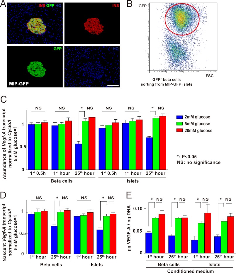
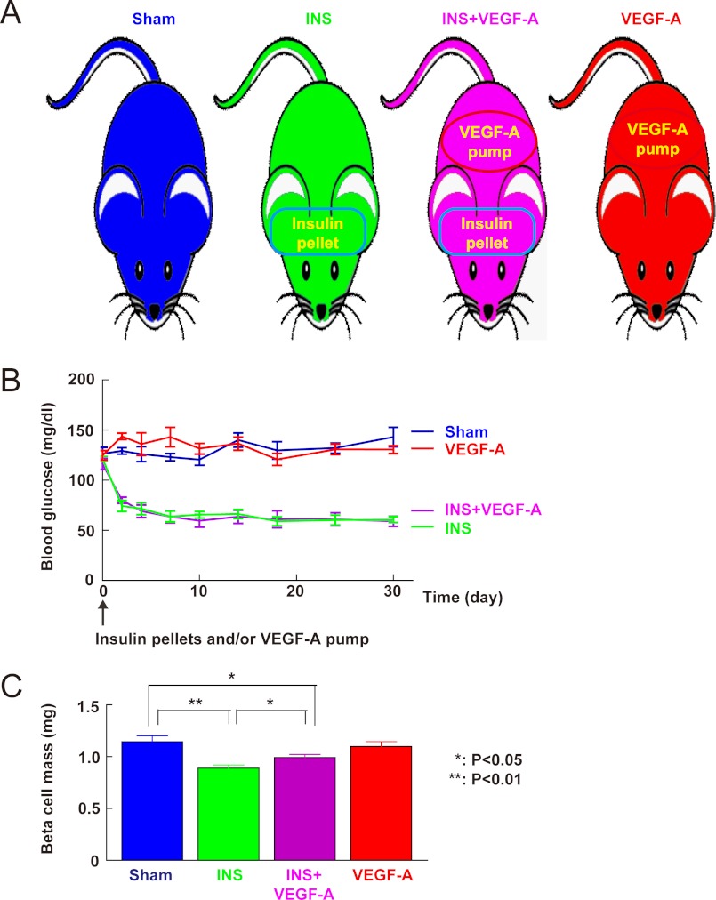
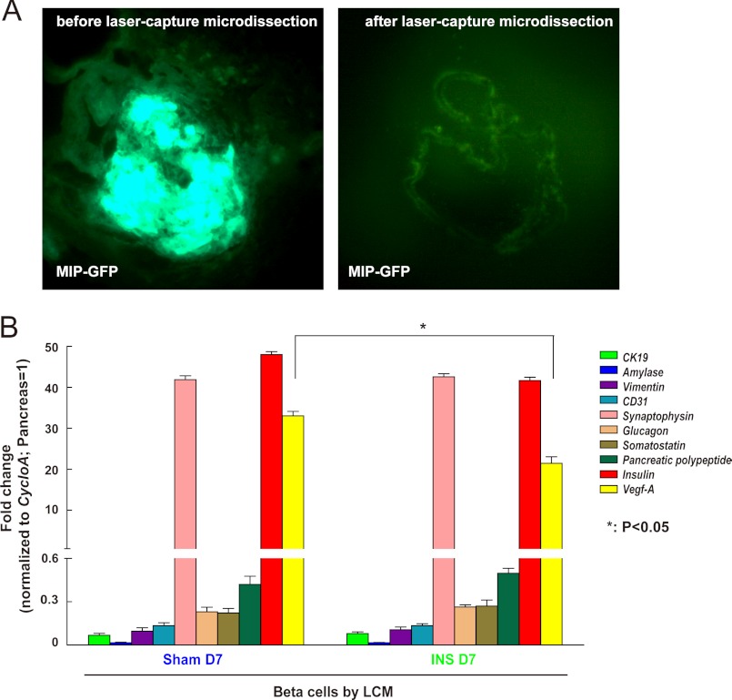
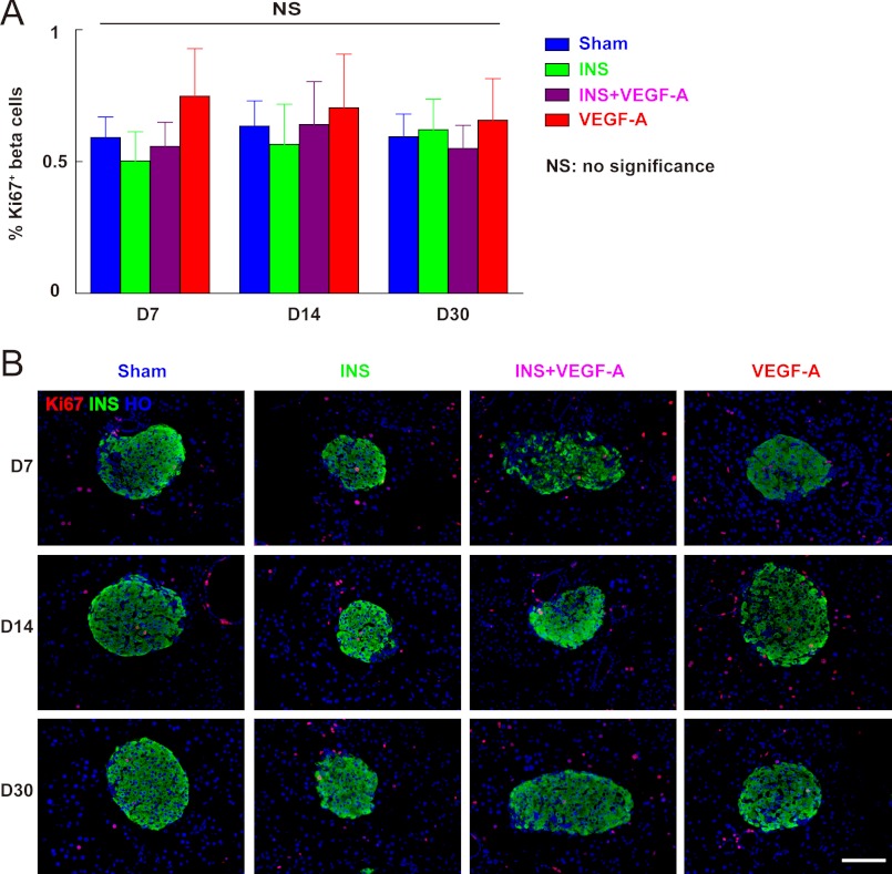
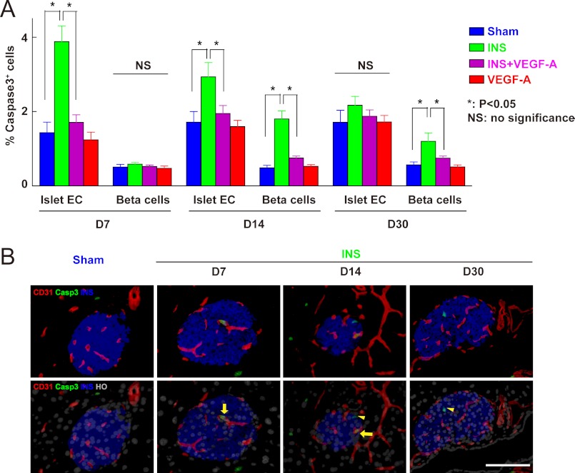
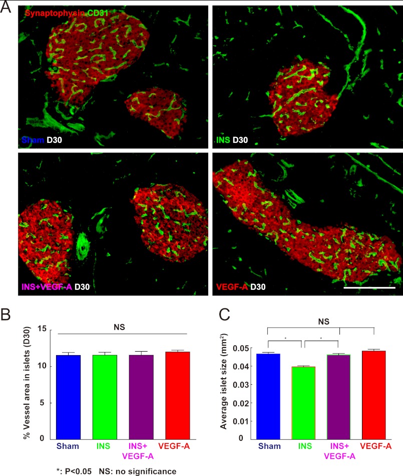
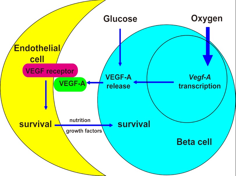
VEGF-A expression in beta cells is critical for pancreatic development, formation of islet-specific vasculature, and Insulin secretion. However, two key questions remain. First, is VEGF-A release from beta cells coupled to VEGF-A production in beta cells? Second, how is the VEGF-A response by beta cells affected by metabolic signals? Here, we show that VEGF-A secretion, but not gene transcription, in either cultured islets or purified pancreatic beta cells, was significantly reduced early on during low glucose conditions. In vivo, a sustained hypoglycemia in mice was induced with Insulin pellets, resulting in a significant reduction in beta cell mass. This loss of beta cell mass could be significantly rescued with continuous delivery of exogenous VEGF-A, which had no effect on beta cell mass in normoglycemic mice. In addition, an increase in apoptotic endothelial cells during hypoglycemia preceded an increase in apoptotic beta cells. Both endothelial and beta cell apoptosis were prevented by exogenous VEGF-A, suggesting a possible causative relationship between reduced VEGF-A and the loss of islet vasculature and beta cells. Furthermore, in none of these experimental groups did beta cell proliferation and islet vessel density change, suggesting a tightly regulated balance between these two cellular compartments. The average islet size decreased in hypoglycemia, which was also prevented by exogenous VEGF-A. Taken together, our data suggest that VEGF-A release in beta cells is independent of VEGF-A synthesis. Beta cell mass can be regulated through modulated release of VEGF-A from beta cells based on physiological need.


Figures

FIGURE 1.
FIGURE 1.
VEGF-A release, but not transcription, from islets or beta cells was reduced in early low glucose culture. A, representative image of a histologic section from a MIP-GFP mouse pancreas showing co-localization of insulin (INS, red) and GFP. B, FACS of MIP-GFP mouse islets: beta cells were isolated based on green fluorescence. C-E, to examine whether VEGF-A release in beta cells is separate from VEGF-A synthesis, we analyzed the release and gene transcription of Vegf-A by either isolated islets or re-aggregated beta cells at 0.5, 1, and 25 h (for the latter, fresh medium was added at 24 h) in serum-free medium supplemented with 2, 5, or 20 mm glucose. C, quantitative RT-PCR was performed to check Vegf-A transcripts in cultured islets or beta cells. Cyclophilin A (cycloA) was used as a housekeeping gene to normalize Vegf-A values. Exposure to high glucose did not change the levels of Vegf-A transcript, whereas exposure to low glucose did not change the levels of Vegf-A transcript within 1 h, but did so at 25 h. D, nuclear run-on assay was performed on cultured islets or beta cells and showed that nascent Vegf-A transcription did not change within the 1-h exposure to low glucose. E, total DNA content of the cells was used to normalize the quantity of released VEGF-A into culture medium. VEGF-A release by either islets or beta cells was significantly reduced in 2 mm glucose (p < 0.05) at both 1 and 25 h. However, 20 mm glucose did not significantly increase VEGF-A release. *, p < 0.05; **, p < 0.01; NS, no significance; INS, insulin; HO, Hoechst. Scale bars are 30 μm.
FIGURE 2.
FIGURE 2.
Reduction of beta cell mass during sustained hypoglycemia can be partially rescued by exogenous VEGF-A. A, experimental design. To evaluate whether hypoglycemia has an effect on beta cell mass, insulin pellets were implanted subcutaneously in mice to induce sustained hypoglycemia (INS, green). To check whether any effect of hypoglycemia on beta cell mass is due to the reduced VEGF-A, insulin pellet-treated mice were rescued with an additionally implanted VEGF-A-releasing pump (INS + VEGF-A, purple). To exclude an independent effect of VEGF-A on beta cell mass, treatment with exogenous VEGF-A alone (VEGF-A, red) was included as a control. The mice from both sham (Sham, blue) and INS groups also received a sham operation to control for the effect of surgery. B, non-fasting blood glucose showed sustained hypoglycemia in mice that received either insulin only (INS, green) or a combination of insulin and VEGF-A releasing pumps (INS + VEGF-A, purple). Mice that received VEGF-A pumps only (VEGF-A, red) were normoglycemic like sham-treated mice (Sham, blue). C, beta cell mass analysis showed a reduction (p < 0.01) in insulin-treated mice (INS, green) compared with sham-treated mice (Sham, blue). This reduction of beta cell mass was significantly attenuated (p < 0.05) in the insulin-treated mice that also received VEGF-A pumps (INS + VEGF-A, purple). VEGF-A pump (VEGF-A, red) itself did not affect (no significance) beta cell mass in normoglycemic mouse controls. *, p < 0.05; **, p < 0.01.
FIGURE 3.
FIGURE 3.
Vegf-A transcripts decrease in beta cells during sustained hypoglycemia. A, representative images before and after beta cell isolation from MIP-GFP mice by LCM. B, levels of mRNA for CK19, Amylase, Vimentin, CD31, Synaptophysin, Glucagon, Somatostatin, Pancreatic polypeptide, and Insulin were evaluated in the isolated beta cells by LCM to assure the purity of the beta cells. Gene values are normalized with cyclophilin A and compared with levels from total pancreas. The Vegf-A transcripts significantly decreased in the beta cells from the hypoglycemic mice. *, p < 0.05.
FIGURE 4.
FIGURE 4.
Beta cell proliferation was not affected by hypoglycemia regardless of exogenous VEGF-A supply. The percentage of pancreatic beta cells that are positive for Ki-67, a cellular marker for proliferation, was quantified at 7, 14, and 30 days. A, Ki-67+ beta cell percentages across all four experimental conditions were consistent (no difference). B, representative immunofluorescent images of mouse pancreas at day 7 (D7), 14 (D14), and 30 (D30) are shown: insulin (INS) in green; Ki-67 in red, and Hoechst (HO) in blue. NS, no significance. Scale bars are 50 μm.
FIGURE 5.
FIGURE 5.
Apoptosis of islet endothelial cells precedes apoptosis of beta cells during sustained hypoglycemia, and can be prevented with exogenous VEGF-A. A-C, pancreases from mice treated with insulin pellets (INS, green), combined insulin pellets and VEGF-A pump (INS + VEGF-A, purple), untreated sham controls (Sham, blue), and VEGF-A only controls (VEGF-A, red) were analyzed at 7, 14, and 30 days for apoptotic islet endothelial cells and beta cells. A, the percentage of islet vessels that contained caspase 3+ endothelial cells (CD31+) was quantified and showed an increase (p < 0.05) at days 7 and 14, but reverted to normal at day 30 after insulin pellet treatment. This apoptosis of endothelial cells can be completely prevented by exogenous VEGF-A. The percentage of caspase 3+ beta cells was also quantified and showed no change at day 7 after insulin pellet treatment, compared with controls. However, this percentage increased by day 14. Similarly, beta cell apoptosis was significantly reduced by exogenous VEGF-A. B, representative immunofluorescent images of mouse pancreas from different time points (D7, D14, and D30) after insulin pellet treatments are shown: CD31 in red, insulin (INS) in blue, caspase 3 in green, and nuclei staining (Hoechst, HO) in gray. Arrows point to caspase 3+ CD31+ cells and arrowheads point to caspase 3+ INS+ cells. Casp3, caspase 3; EC, endothelial cell. *, p < 0.05; NS, no significance. Scale bars are 50 μm.
FIGURE 6.
FIGURE 6.
Quantification of islet vessel density and average islet size. A and B, islet vessel densities were measured under all experimental conditions and evaluated with the ratio of islet CD31+ cell area to synaptophysin+ cell area. Synaptophysin is a pan-endocrine cell marker. A, representative immunofluorescent images of mouse pancreas at day 30 were shown: CD31 in green and synaptophysin in red. B, the islet vessel densities were consistent (no difference) across all four experimental conditions. C, average islet size was measured among all four experimental conditions, showing a significant decrease in insulin-treated mice, which was prevented with exogenous VEGF-A. *, p < 0.05; NS, no significance. Scale bars are 50 μm.
FIGURE 7.
FIGURE 7.
Hypoglycemia regulates beta cell mass via modulated VEGF-A release. A model of how beta cell mass is regulated by hypoglycemia via VEGF-A is proposed. Oxygen tension is the major regulator for Vegf-A transcription in pancreatic beta cells. Hypoxia can greatly increase Vegf-A transcription. However, glucose can regulate VEGF-A release from beta cells before the adaptation of Vegf-A transcription occurs. Hypoglycemia can reduce VEGF-A release, resulting in a decrease in survival of neighboring islet endothelial cells, which subsequently leads to a secondary loss of beta cells.

References

    1. Cleaver O., Melton D. A. (2003) Endothelial signaling during development. Nat. Med. 9, 661–668 - PubMed
    1. Ferrara N., Gerber H. P., LeCouter J. (2003) The biology of VEGF and its receptors. Nat. Med. 9, 669–676 - PubMed
    1. Carmeliet P., Jain R. K. (2011) Molecular mechanisms and clinical applications of angiogenesis. Nature 473, 298–307 - PMC - PubMed
    1. Lee S., Jilani S. M., Nikolova G. V., Carpizo D., Iruela-Arispe M. L. (2005) Processing of VEGF-A by matrix metalloproteinases regulates bioavailability and vascular patterning in tumors. J. Cell Biol. 169, 681–691 - PMC - PubMed
    1. Stalmans I., Ng Y. S., Rohan R., Fruttiger M., Bouché A., Yuce A., Fujisawa H., Hermans B., Shani M., Jansen S., Hicklin D., Anderson D. J., Gardiner T., Hammes H. P., Moons L., Dewerchin M., Collen D., Carmeliet P., D'Amore P. A. (2002) Arteriolar and venular patterning in retinas of mice selectively expressing VEGF isoforms. J. Clin. Invest. 109, 327–336 - PMC - PubMed

Publication types

MeSH terms

Substances