. 2013 Mar 22;288(12):8726-8736.
doi: 10.1074/jbc.M112.368829. Epub 2013 Feb 1.

RNF13, a RING finger protein, mediates endoplasmic reticulum stress-induced apoptosis through the inositol-requiring enzyme (IRE1α)/c-Jun NH2-terminal kinase pathway

Affiliations

RNF13, a RING finger protein, mediates endoplasmic reticulum stress-induced apoptosis through the inositol-requiring enzyme (IRE1α)/c-Jun NH2-terminal kinase pathway

Muhammad Arshad et al. J Biol Chem. .

Abstract

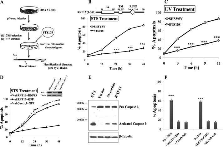
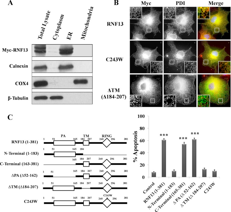
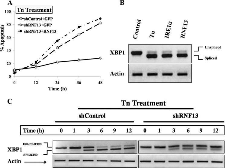
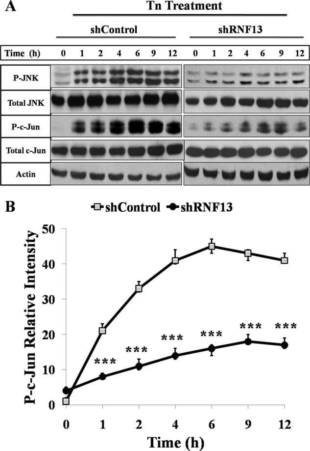
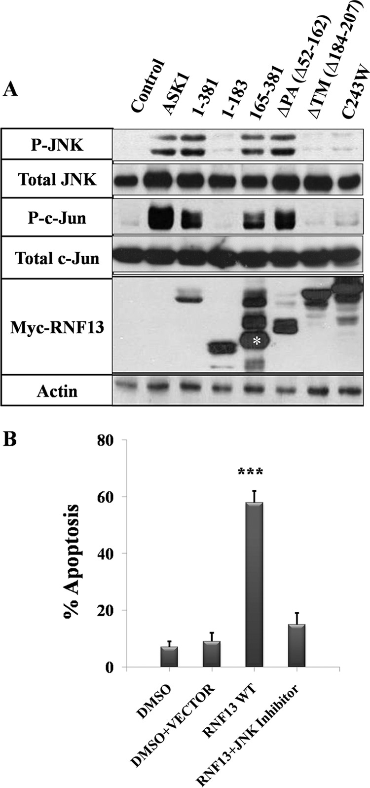
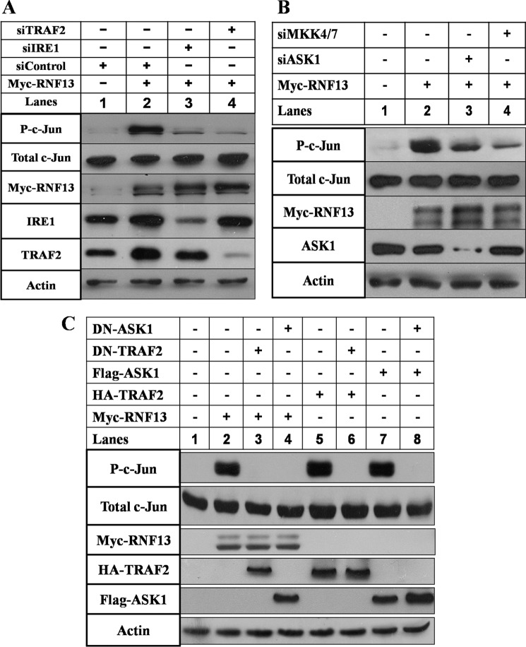
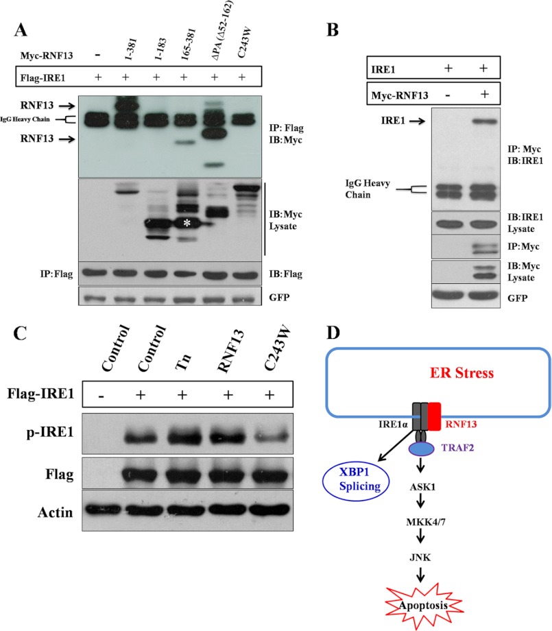
Disturbance of homeostasis at endoplasmic reticulum (ER) causes stress to cells that in turn triggers an adaptive signaling pathway termed unfolded protein response for the purpose of restoring normal cellular physiology or initiating signaling events leading to apoptosis. Identification of those genes that are involved in the unfolded protein response-mediated apoptotic signaling pathway would be valuable toward elucidating the molecular mechanism underlying the relationship between ER stress and apoptosis. We initiated a genetic screen by using the retroviral insertion mutation system to search for genes whose inactivation confers resistance to apoptosis induction by staurosporine. Using this approach, RING finger protein 13 (RNF13) was identified. Interestingly, RNF13 is highly enriched in ER. RNF13 knockdown cells are resistant to apoptosis and JNK activation triggered by ER stress. Conversely, overexpression of RNF13 induces JNK activation and caspase-dependent apoptosis. The RING and transmembrane domains of RNF13 are both required for its effects on JNK activation and apoptosis. Moreover, systematic analysis of the involvement of individual signaling components in the ER stress pathway using knockdown approach reveals that RNF13 acts upstream of the IRE1α-TRAF2 signaling axis for JNK activation and apoptosis. Finally, RNF13 co-immunoprecipitates with IRE1α, and the intact RING domain is also required for mediating its interaction. Together, our data support a model that RNF13 is a critical mediator for facilitating ER stress-induced apoptosis through the activation of the IRE1α-TRAF2-JNK signaling pathway.


Figures

FIGURE 1.
FIGURE 1.
Identification of RNF13 as a novel apoptotic regulator. A, a schematic diagram depicts procedures for conducting the retroviral random insertion screen. B, top, a schematic illustrates domain organization in RNF13. The predicted full-length RNF13 protein consists of a protease-associated domain (PA) at the N terminus, a transmembrane domain (TM) in the middle, and a really interesting new gene containing domain (RING) at the C terminus as indicated. Bottom, STS-108 is a SHSY-5Y cell clone identified to be STS-resistant during the screening. Wild-type and STS-108 SHSY-5Y cells were treated with 1 μm STS for the indicated period of time. The nucleus was visualized by staining with 100 ng/ml Hoechst 33342, and cells containing the condensed or broken nuclei were defined as apoptotic cells under fluorescence microscope. 300 cells were chosen randomly for scoring, and the percentage of apoptotic cells was calculated by the number of apoptotic cells over total number of cells. C, STS-108 is also resistant to UV-induced apoptosis. Wild-type and STS-108 SHSY-5Y cells were exposed to 10 mJ/cm2 of UV radiation and incubated for the indicated periods of time before they were examined for apoptosis as described in B. D, reintroduction of siRNA-resistant Myc-tagged RNF13 into RNF13 knockdown (shRNF13) cells restores their sensitivity to STS-induced apoptosis. Wild-type and RNF13 knockdown SHSY-5Y cells were co-transfected with 0.2 μg of GFP and 3 μg of CMV5 or Myc-RNF13 plasmid for 36 h. Cells were then treated with 1 μm STS for the indicated amounts of time and subsequently evaluated for apoptosis. The inset panel is the result of RT-PCR for RNF13 mRNA levels. E, overexpression of RNF13 induces caspase-3 activation. COS-7 cells were treated with 1 μm STS for 3 h or transfected with 3 μg of indicated plasmids for 30 h. Cell lysates were blotted with anti-caspase-3 antibody. M-cofilin is the mitochondrial-targeted cofilin that induces caspase activation and apoptosis as shown previously (20). F, RNF13 induces caspase-dependent apoptosis. COS-7 cells transfected with 3 μg of plasmid encoding M-cofilin or RNF13 in the presence or absence of indicated caspase inhibitors were analyzed. At 36 h of post-transfection, cells were evaluated for apoptosis as in B. Data are representative of three independent experiments. ZVAD-fmk, benzyloxycarbonyl-VAD-fluoromethyl ketone. ***, p < 0.001.
FIGURE 2.
FIGURE 2.
RNF13 is highly enriched in the microsomal ER fraction, and both the ER localization and functional RING domain are required for RNF13-induced apoptosis. A, mitochondrial, microsomal ER, and cytosolic fractions of 293T cells transfected with Myc-RNF13 were prepared as described under “Materials and Methods.” Different components were subjected to SDS-PAGE and blotted with the corresponding antibodies as indicated. B, the putative TM of RNF13 is required for mediating its localization to ER. Myc-tagged RNF13 and its mutations were expressed in COS-7 cells. Immunostaining was performed as described under “Materials and Methods”; ER was stained with anti-protein-disulfide isomerase (PDI) antibody. Insets show enlargements of the boxed regions. C, the TM and the intact RING domains of RNF13 are both required for mediating its proapoptotic activity. Left, shown is a schematic representation of RNF13 and mutants used in experiments. Right, COS-7 cells were transfected with either 3 μg of plasmid encoding wild-type RNF13 or the indicated mutants for 36 h before the evaluation of apoptosis. Data are representative of three independent experiments. ***, p < 0.001.
FIGURE 3.
FIGURE 3.
RNF13 is required for ER stress-induced apoptosis. A, RNF13 knockdown SHSY-5Y cells are resistant to Tn-induced apoptosis, and introduction of the expression plasmid encoding the siRNA-resistant RNF13 cDNA into the knockdown cells restores its sensitivities to Tn. Wild-type and RNF13 knockdown SHSY-5Y cells were co-transfected with 0.2 μg of GFP plasmid and 3 μg of CMV5 or siRNF13–1-resistant Myc-RNF13 plasmid for 36 h. Cells were then treated with Tn (1.25 μg/ml) for the indicated amount of time before apoptosis assay was performed. B, overexpression of RNF13 induces XBP1 splicing. SHSY-5Y cells were treated with 1.25 μg/ml Tn for 6 h or transfected with 3 μg of either IRE1α or RNF13 plasmid for 24 h. Cells were harvested, RNA was extracted, and the levels of unspliced and spliced XBP1 were evaluated by RT-PCR analysis. β-Actin was used as a loading control. C, Tn-induced splicing of XBP1 mRNA is attenuated in the RNF13 knockdown SHSY-5Y cells. Control and RNF13 knockdown SHSY-5Y cells were treated with 1.25 μg/ml Tn for indicated periods of time before harvesting for RNA extraction. RT-PCR analysis was then performed to observe subsequent XBP1 splicing. β-Actin was served as a loading control.
FIGURE 4.
FIGURE 4.
Activation of JNK and phosphorylation of c-Jun by Tn is suppressed in the RNF13 knockdown cells. A, SHSY-5Y cells were treated with 1.25 μg/ml Tn for indicated amounts of time before harvesting for Western blot analysis. Western blot analysis of the levels of phosphorylated endogenous JNK and c-Jun in wild-type and RNF13 knockdown cells is shown. B, shown is quantitative analysis of the relative levels of phosphorylated c-Jun in A using Quantity One software. β-Actin was used as a loading control. Data presented are the mean ± S.D. from three independent experiments. ***, p < 0.001.
FIGURE 5.
FIGURE 5.
The RING and TM domains are both required for RNF13-induced JNK activation. A, COS-7 cells were transfected with 3 μg of plasmid encoding the Myc-tagged RNF13 or the indicated mutant for 30 h. Cells were harvested, and a Western blot was performed using antibodies against phospho-JNK and phospho-c-Jun. β-Actin was used as a loading control. The asterisk represents the correct sized protein band for RNF13 truncation (aa 165–381). B, inhibition of JNK activity blocks RNF13-induced apoptosis. COS-7 cells preincubated with 10 μm JNK inhibitor (SP600125) were transfected with 3 μg of Myc-tagged RNF13 for 30 h before apoptosis of was analyzed. Data presented are the mean ± S.D. values from three independent experiments. ***, p < 0.001.
FIGURE 6.
FIGURE 6.
RNF13 induces JNK activation via the IRE1α-TRAF2-ASK1-MKK4/7-JNK signaling axis. A, knockdown of IRE1α or TRAF2 blocks RNF13-induced c-Jun phosphorylation. SHSY-5Y cells were co-transfected with 3 μg of plasmid of RNF13 and 3 μg of indicated siRNAs corresponding to IRE1α and TRAF2 for 30 h before harvesting for Western blot analysis. Western blotting was performed using antibodies against c-Jun, phosphorylated c-Jun, Myc, IRE1α, and TRAF2 as indicated on the left. β-Actin served as a loading control. B, activation of c-Jun by RNF13 is dependent on ASK1 and MKK4/7. Knockdown of ASK1 or MKK4/7 inhibits RNF13-mediated c-Jun phosphorylation in SHSY-5Y cells. Cells were co-transfected with 3 μg of RNF13 and the indicated siRNAs for 30 h. Cells were harvested, and Western blot analysis was performed using antibodies against c-Jun, phospho-c-Jun, Myc, and ASK1. C, expression of the dominant negative (DN) form of TRAF2 or ASK1 inhibits RNF13-mediated c-Jun activation. SHSY-5Y cells were co-transfected with 3 μg of the indicated plasmids and plasmid encoding the indicated dominant negative mutant for 30 h before harvesting for Western blots. Antibodies against c-Jun, phospho-c-Jun, Myc, HA, and FLAG were used to measure the amounts of c-Jun, phosphorylated c-Jun, RNF13, TRAF2, and ASK1. β-Actin was used as a loading control.
FIGURE 7.
FIGURE 7.
RNF13 interacts with IRE1α and promotes IRE1α phosphorylation. A, 293T cells were co-transfected with 2 μg of FLAG-IRE1α plasmid and 3 μg of plasmid encoding Myc-RNF13 or its mutants for 24 h. FLAG-IRE1α protein was immune-precipitated (IP) using anti-FLAG antibody, and the presence of RNF13 or its mutant proteins in the immunoprecipitated products was detected by anti-Myc antibody. IB, immunoblot. B, RNF13 interacts with endogenous IRE1α. 293T cells were transfected with 3 μg Myc-RNF13 plasmid for 24 h. Myc-RNF13 protein was immunoprecipitated using anti-Myc antibody, and the presence of endogenous IRE1α in the immunoprecipitated products was detected by anti-IRE1α antibody. C, 293T cells were transfected with 2 μg of IRE1α plasmid alone or co-transfected with 3 μg of plasmid encoding RNF13 or its RING domain mutant (C243W) for 24 h. For cells treated with Tn, 1.25 μg/ml drug was added at 18 h post-transfection. D, a schematic model illustrates the role of RNF13 in mediating apoptosis through the IRE1α-TRAF2-JNK signaling pathway.

References

    1. Kim I., Xu W., Reed J. C. (2008) Cell death and endoplasmic reticulum stress. Disease relevance and therapeutic opportunities. Nat. Rev. Drug Discov. 7, 1013–1030 - PubMed
    1. Walter P., Ron D. (2011) The unfolded protein response. From stress pathway to homeostatic regulation. Science 334, 1081–1086 - PubMed
    1. Cnop M., Foufelle F., Velloso L. A. (2012) Endoplasmic reticulum stress, obesity, and diabetes. Trends Mol. Med. 18, 59–68 - PubMed
    1. Merquiol E., Uzi D., Mueller T., Goldenberg D., Nahmias Y., Xavier R. J., Tirosh B., Shibolet O. (2011) HCV causes chronic endoplasmic reticulum stress leading to adaptation and interference with the unfolded protein response. Plos One 6, e24660. - PMC - PubMed
    1. Malhotra J. D., Kaufman R. J. (2007) The endoplasmic reticulum and the unfolded protein response. Semin. Cell Dev. Biol. 18, 716–731 - PMC - PubMed

Publication types

MeSH terms