Review
. 2013 Feb 1;5(2):a008730.
doi: 10.1101/cshperspect.a008730.

Inhibitor of apoptosis (IAP) proteins-modulators of cell death and inflammation

Affiliations
Review

Inhibitor of apoptosis (IAP) proteins-modulators of cell death and inflammation

John Silke et al. Cold Spring Harb Perspect Biol. .

Abstract

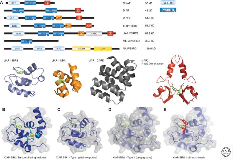
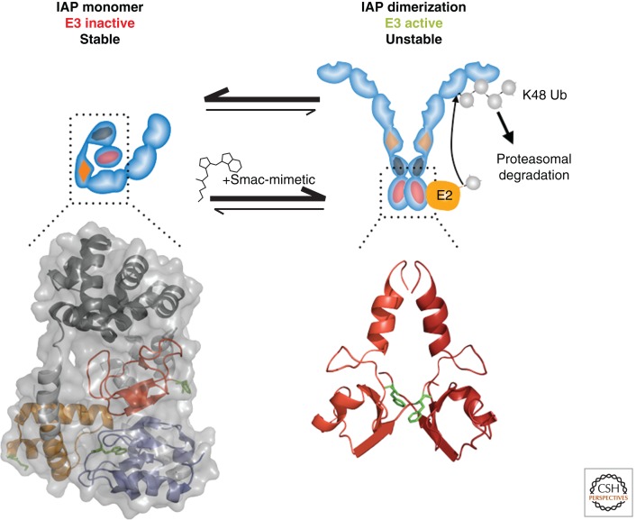
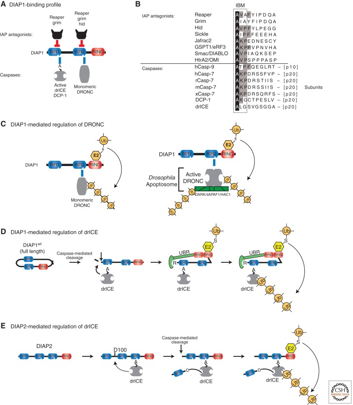
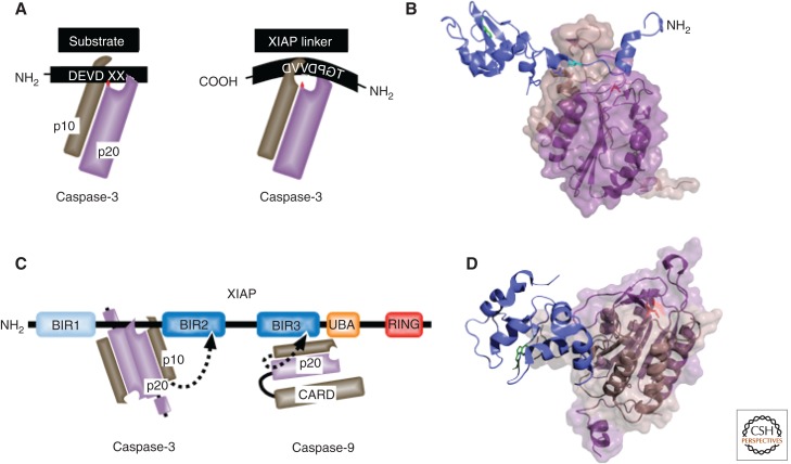
Misregulated innate immune signaling and cell death form the basis of much human disease pathogenesis. Inhibitor of apoptosis (IAP) protein family members are frequently overexpressed in cancer and contribute to tumor cell survival, chemo-resistance, disease progression, and poor prognosis. Although best known for their ability to regulate caspases, IAPs also influence ubiquitin (Ub)-dependent pathways that modulate innate immune signaling via activation of nuclear factor κB (NF-κB). Recent research into IAP biology has unearthed unexpected roles for this group of proteins. In addition, the advances in our understanding of the molecular mechanisms that IAPs use to regulate cell death and innate immune responses have provided new insights into disease states and suggested novel intervention strategies. Here we review the functions assigned to those IAP proteins that act at the intersection of cell death regulation and inflammatory signaling.


Figures

Figure 1.
Figure 1.
Domain architecture of IAPs. The first IAP (OpIAP) was identified from a baculovirus strain in 1993 by Miller and colleagues, based on its ability to suppress virus-induced apoptosis of infected cells. Cellular IAPs were subsequently identified in insects and vertebrates. (A) Schematic and domain structures of some of the IAPs discussed in this review. All schematic IAPs, except NAIP, are drawn to scale, Leucine-rich repeats (LRR) and NACHT, the domain present in NAIP, CIITA, HET-E, and TP1. BIR domains provide interactions with proteins such as caspases, IAP-antagonists, TRAF1/2, and TAB1. cIAP1’s BIR3 is represented as a blue cartoon structure, and a highly conserved Trp in the IBM binding pocket is represented in green stick format. The UBA domain of cIAP1 binds to poly-Ub and is represented as an orange cartoon structure with a highly conserved Met represented in green stick format as a reference point. The caspase recruitment domain (CARD) present in cIAPs, which generally serves as a protein-interaction surface, has an unknown function in IAPs, and the CARD of cIAP1 is represented as a grey cartoon structure. The carboxy-terminal really interesting new gene (RING) domain is required for Ub-ligase activity and serves as dimerization interface and docking site for E2s. A dimeric cIAP2 structure is represented as a cartoon structure with each monomer in a different shade of red. A highly conserved Phe is represented in green stick format. Coordinates are from Dueber and colleagues (Dueber et al. 2011), PDB: 3T6P, and Mace and colleagues (Mace et al. 2008), PDB: 3EB5. (B) XIAP BIR3 with Zn ion (yellow sphere) and Zn coordinating cysteines and histidine are represented in cyan. Coordinates are from Mastrangelo and colleagues (Mastrangelo et al. 2008), PDB: 3CM2. (C) The BIR1 of XIAP (Type I) is represented as a cartoon and transparent surface, conserved Trp is indicated in green stick format. Coordinates are from Lu and colleagues (Lu et al. 2001), PDB: 2POI. (D) XIAP BIR3 (Type II). A conserved Trp residue in the IBM groove is represented in green stick format. (E) XIAP BIR3 bound to a Smac peptide is represented in red stick format. All crystal structure pictures were generated with PyMOL.
Figure 2.
Figure 2.
Mechanism of Smac-mimetic (SM)-induced activation of cIAPs. Monomeric cIAP1 BIR3, UBA, CARD RING, represented as in Fig. 1 using coordinates from Dueber and colleagues (Dueber et al. 2011), PDB: 3T6P, is an inactive E3 ligase. Binding of an SM releases the BIR3-mediated inhibition on RING dimerization (PDB:3EB5), resulting in activation of the E3 ligase function, auto-K48 ubiquitylation, and proteasomal degradation.
Figure 3.
Figure 3.
IAP-mediated regulation of caspases in Drosophila. (A) Binding profile of DIAP1 with caspases and IAP antagonists. Direct physical interaction with the effector caspases drICE or DCP-1 and the initiator caspase DRONC is mediated through DIAP1’s BIR1 and BIR2 domains, respectively. Following their activation, drICE and DCP-1 expose an NH2-terminal IBM (depicted as A), which allows their binding to BIR1. (B) Sequence alignment of IBM-bearing proteins. Identical residues are highlighted in black. Residues conserved in four or more IBM proteins are indicated in gray. (C) DIAP1’s BIR2-DRONC association is essential for DIAP1 to neutralize DRONC. Following binding, DIAP’s RING finger promotes Ub conjugation of DRONC, leading to its inactivation through nondegradative ubiquitylation of monomeric DRONC (left panel) and by targeting apoptosome-associated active DRONC for degradation (right panel). (D,E) Mechanism of effector caspase (drICE) inactivation by DIAP1 (D) and DIAP2 (E). (D) Full-length wild-type DIAP1 is held in an inactive conformation and requires caspase-mediated proteolytic cleavage at residue 20 for its activation. After cleavage, BIR-mediated caspase binding occurs more efficiently. Cleavage also facilitates recruitment of N-end rule UBR E3 ligases, which together with DIAP1’s RING domain, promote ubiquitylation and inactivation of drICE and DCP-1. (E) drICE is also subject to regulation by DIAP2. drICE binds to the BIR3 of DIAP2 in an IBM-dependent manner and, following binding, cleaves DIAP2 at D100. DIAP2 cleavage results in a covalent adduct between D100 and the catalytic machinery of drICE, trapping the caspase. Full inactivation of drICE is achieved through RING-mediated ubiquitylation.
Figure 4.
Figure 4.
XIAP-mediated inhibition of caspase-3 and caspase-9. (A) Schematic comparison of substrate and XIAP linker interaction with caspase-3, catalytic cysteine, indicated in red. (B) The BIR2 of XIAP and NH2-terminal linker (blue cartoon) embedded in the active site groove of the caspase-3 p10 (brown)/p20 (violet) heterodimer (PDB: 1I30), revealing the reverse (C-N) linker, cleavage incompatible, orientation. The reference Trp in the IBM binding groove of XIAP’s BIR2 is indicated in green, Asp148 in cyan, and the position of the catalytically active cysteine in the p20 of caspase-3 in red stick format. (C) The distinct mechanisms of caspase inhibition used by XIAP represented schematically. The BIR3 binds to a dimerization surface of a caspase-9 monomer, preventing it from dimerizing and autoactivating. (D) The structure (PDB: 1NW9) of the BIR3 of XIAP (blue) bound to caspase-9 (p10 in brown, p20 in violet). The conserved reference Trp in the IBM binding groove of BIR3 is indicated in green stick format and the position of the catalytic cysteine in red stick format.
Figure 5.
Figure 5.
cIAP1, cIAP2, and XIAP prevent the formation of a RIPK1-dependent platform, dubbed the Ripoptosome, Necrosome, or Complex-II. (A) All three IAPs target RIPK1 and components of the Ripoptosome (caspase-8 and cFLIPL) for Ub-mediated inactivation. Following genotoxic stress, cytokine signaling-induced depletion of cIAPs, or SM treatment, cIAP1, cIAP2, and XIAP levels rapidly decline and/or are inactivated. This allows formation and accumulation of the Ripoptosome. In the presence of high levels of RIPK3, this can lead to necroptosis. cFLIP also regulates Ripoptosome-mediated cell death. cFLIPL thereby prevents apoptosis and necroptosis, whereas FLIPS inhibits apoptosis but promotes necroptosis. (B) Under steady-state conditions, the majority of RIPK1 appears to be in a closed configuration that prevents it from binding to partner proteins. Cytokine receptor stimulation can convert a small fraction of RIPK1 into an “open,” binding-competent configuration. In the presence of cIAPs and XIAP, binding-competent RIPK1 is targeted for Ub-mediated inactivation, most likely via proteasomal degradation. Under conditions where IAP levels are low, however, unmodified and binding-competent RIPK1 accumulates and can form the Ripoptosome. In the presence of high levels of cFLIPL, the Ripoptosome is dissolved via caspase-8-cFLIPL-mediated cleavage of RIPK1. When cFLIPL levels are low, the Ripoptosome can promote caspase-dependent or caspase-independent cell death.
Figure 6.
Figure 6.
TNF signaling is regulated by cIAPs. (A) Upon TNF-binding, cIAPs are recruited to the TNF-R1 signaling complex (Complex-I) via TRADD/TRAF2. The cIAP RING dimerizes, which leads to activation of its E3 activity. Active cIAPs ubiquitylate several molecules within the complex. RIPK1 ubiquitylation is the most readily observed, and is absent in SM-treated or cIAP knockout cells. Of note, although TNF-induced ubiquitylation of RIPK1 is a prominent event, RIPK1 is not required for TNF signaling in all cells. Ubiquitylation of components of Complex-I, such as RIP, drives the recruitment of HOIL-1/HOIP/Sharpin that together form the linear ubiquitin assembly complex (LUBAC). LUBAC generates linear Ub chains on NEMO and RIPK1, which in turn recruits more NEMO molecules via its linear Ub-binding UBAN domain. NEMO is probably constitutively associated with IKKα/IKKβ, and IKKβ is phosphorylated and activated by TAK1 that is independently recruited to ubiquitylated Complex-I via its Ub receptors TAB2 and TAB3, which bind only to K63-linked Ub chains. Phosphorylated and activated IKKβ in turn phosphorylates IκBα, which leads to recruitment of a HECT E3 ligase. This E3 ligase promotes K48-linked ubiquitylation and proteasomal degradation of IκBα, allowing translocation of NF-κB subunits p50 and p65 to drive production of cytokines. p50 and p65 also promote expression of IκBα to cause feedback inhibition, as well as genes such as cFLIP that are required to protect cells from Complex-II-induced cell death. The numbered arrows provide a tentative indication of temporal sequence. Complex-II is most likely generated from Complex-I, in an as yet undefined manner, and comprises FADD and caspase-8. cIAPs and LUBAC appear to limit Complex-II formation by promoting ubiquitylation-mediated degradation of Complex-II components. Caspase-8 limits Complex-II formation by cleaving and inactivating RIPK1. Therefore, loss of IAPs, LUBAC, or caspase-8 activity results in formation of Complex-II*, which is able to drive necroptosis. (B) Schematic diagram depicting NOD-mediated signaling. Upon stimulation of NOD1 or NOD2 by their respective ligands (DAP and MDP), a similar signaling complex to that of TNF-R1 is assembled. However, whereas cIAPs are critical regulators of TNF-R1 signaling, XIAP plays a key role in NOD-mediated activation of NF-κB and MAPK. XIAP, thereby, allows signaling by targeting NOD-bound RIPK2 for ubiquitylation.

References

    1. Arama E, Agapite J, Steller H 2003. Caspase activity and a specific cytochrome C are required for sperm differentiation in Drosophila. Dev Cell 4: 687–697 - PubMed
    1. Bartke T, Pohl C, Pyrowolakis G, Jentsch S 2004. Dual role of BRUCE as an antiapoptotic IAP and a chimeric E2/E3 ubiquitin ligase. Mol Cell 14: 801–811 - PubMed
    1. Berger AB, Witte MD, Denault JB, Sadaghiani AM, Sexton KM, Salvesen GS, Bogyo M 2006. Identification of early intermediates of caspase activation using selective inhibitors and activity-based probes. Mol Cell 23: 509–521 - PubMed
    1. Bertrand MJ, Doiron K, Labbe K, Korneluk RG, Barker PA, Saleh M 2009. Cellular inhibitors of apoptosis cIAP1 and cIAP2 are required for innate immunity signaling by the pattern recognition receptors NOD1 and NOD2. Immunity 30: 789–801 - PubMed
    1. Bhoj VG, Chen ZJ 2009. Ubiquitylation in innate and adaptive immunity. Nature 458: 430–437 - PubMed

Publication types

MeSH terms

Substances