Case Reports
. 2012 Jun;6(6):1-9.
doi: 10.3941/jrcr.v6i6.1095. Epub 2012 Jun 1.

Presacral myelolipoma: a case report and review of imaging findings

Affiliations
Case Reports

Presacral myelolipoma: a case report and review of imaging findings

Kevin S Baker et al. J Radiol Case Rep. 2012 Jun.

Abstract

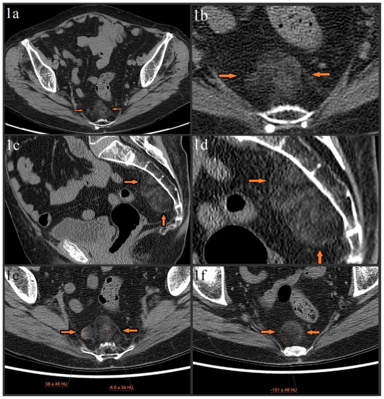
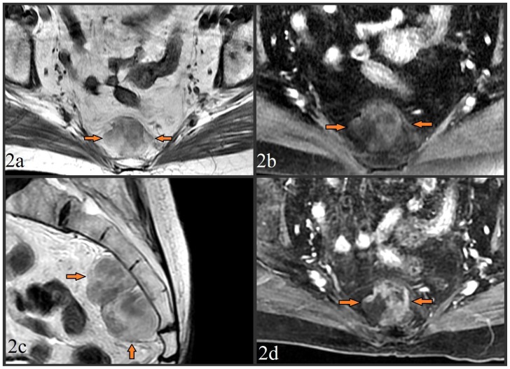
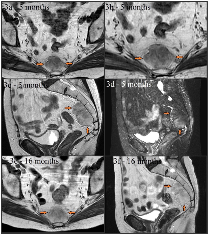
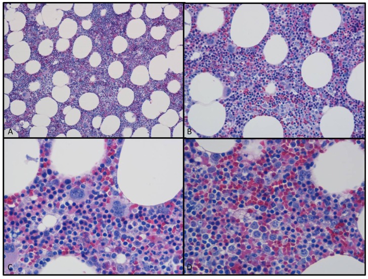
Extra-adrenal myelolipoma is a relatively rare entity, with fewer than 50 cases reported in literature. We present a case of a 79 year-old female who presented for evaluation of hip fracture following trauma, where a lobulated presacral mass with mixed fat/soft tissue attenuation was incidentally seen on initial bone algorithm pelvic CT. Subsequent MRI showed signal characteristics of a lesion with mixed fat and soft tissue composition. The lesion demonstrated stability on follow-up imaging. An elective surgical resection was performed which yielded a grossly fatty mass. The diagnosis of presacral myelolipoma was confirmed on microscopic examination. Following description of our case, we conduct a literature review of the imaging characteristics, diagnosis, and treatment of presacral myelolipoma.

Keywords: MR imaging; Myelolipoma; extra adrenal myelolipoma; lipomatous tumor; presacral CT; presacral MRI; presacral myelolipoma.


Figures

Figure 1
Figure 1
79 year old female with presacral myelolipoma. The CT was performed on a GE® 64-slice CT scanner. All images are from the patient’s initial noncontrast bone algorithm pelvic CT with standard soft tissue windows (width = 400, center = 50). 2mm slices were acquired, using 120 kVp and variable mAs, which ranged from 614 mAs to 633 mAs for the provided axial images. Figures 1a–1f: Axial (figure 1a, magnified in figure 1b) and sagittal (figure 1c, magnified in figure 1d) images show incidental 5.8 × 2.9 × 4.8 cm lobulated, heterogeneous presacral mass without evidence of erosion/invasion of the anterior sacrum (orange arrows). The mass spans from S3 to S5 and is composed of fat and soft tissue elements. Precise measurement of the soft tissue elements’ attenuation is difficult due to significant admixture, but attempts show a range from about −10 HU to about 20 HU, with a single area of higher (mid 30 HUs) attenuation in the lesion’s right lateral margin (image 1e). The fat elements have attenuation values of about −100 HU (image 1f).
Figure 2
Figure 2
MR images from a 79 year old female with presacral myelolipoma. All images were acquired on a GE® 1.5 Tesla MR scanner. Figure 2a. Pre-contrast T1-weighted fast spin echo (FSE) axial image (TR=500.0, TE=9.0, TI=0.0, FA=90.0) at the level of the midsacrum demonstrated a 6.4 × 3.1 × 5.7 cm lobulated presacral mass with mixed fat/soft tissue signal (arrows). No bony invasion was seen. Figure 2b. Pre-contrast fat-suppressed T1-weighted FSE axial image (TR=516.7, TE=7.7, TI=0.0, FA=90.0) taken at the same level, showed loss of signal intensity in the areas which were previously iso-intense to fat, providing further confirmation of a significant fat component of the mass. Figure 2c. Pre-contrast T2-weighted FSE image (TR=5800.0, TE=119.8, TI=0.0, FA=90.0) in the sagittal plane to provide craniocaudad visualization of the 6.4 × 3.1 × 5.7 cm mass which spanned from S3 to S5. The mass was again noted to be lobulated, of mixed fat/soft tissue constituency, and without bony invasion. Figure 2d. Post-contrast (15cc of gadopentetate dimeglumine) T1-weighted fat-suppressed liver acquisition with volume acquisition (LAVA) sequence axial image (TR=4.4, TE=2.1, TI=7.0, FA=12.0) taken at the same level of images 2b/2c showed enhancement of the non-fatty soft tissue elements, and redemonstrated loss of signal in the mass’ fatty elements.
Figure 3
Figure 3
Noncontrast MR images from a 79 year old (80 year old at the time of images 3e and 3f) female with presacral myelolipoma. All images were acquired on a GE® 1.5 Tesla MR scanner. Follow-up axial noncontrast T1-weighted FSE (image 3a and magnified in 3b, TR=400.0, TE=8.5, TI=0.0, FA=90.0), noncontrast sagittal T2-weighted (image 3c, TR=3166.7, TE=122.1, TI=0.0, FA=90.0), and noncontrast sagittal T2-weighted fat-suppressed (image 3d, TR=3450.0, TE=122.1, TI=0.0, FA=90.0) images taken about 5 months later showed stability of the presacral mass (arrows) without significant interval change in size, appearance, or signal characteristics. The mass is again noted to be lobulated, well-circumscribed, and have mixed soft tissue and fat composition (with signal dropout on fat-suppressed image 3d). A lack of sacral invasion is again seen. Noncontrast T1-weighted FSE (image 3e, TR=433.3, TE=7.5, TI=0.0, FA=90.0) and noncontrast sagittal T2-weighted (image 3f, TR=5450.0, TE=119.9, TI=0.0, FA=90.0) images obtained 16 months later also showed continued overall stability.
Figure 4
Figure 4
Photomicrograph from the obtained surgical specimen using H&E (hematoxylin and eosin) staining. Images 4a (10× magnification) and 4b (20× magnification) showed mature adipose tissue with prominent cellular stroma. Images 4c and 4d are higher magnification (both 40x) views which showed that the stroma consisted of all three hematopoietic cell lineages; myeloid, erythroid, and megakaryocytic forming cell lines.

References

    1. Sutker B, Balthazar EJ, Fazzini E. Presacral Myelolipoma: CT findings. J Comput Assist Tomogr. 1985;9:1128–1130. - PubMed
    1. Chen KTK, Felix EL, Flam MS. Extra adrenal myelolipoma. Am J Clin Pathol. 1982 Sep;78(3):386–389. - PubMed
    1. Prahlow IA, Loggie BW, Cappellari JO, Scharling ES, Teot LA, Iskandar SS. Extra-adrenal myelolipoma: report of two cases. South Med J. 1995;88(6):639–643. - PubMed
    1. Grignon DI, Shkrum MJ, Smout MS. Extra-adrenal myelolipoma. Arch Pathol Lab Med. 1989 Jan;113(1):52–54. - PubMed
    1. Kammen BF, Elder DE, Fraker DL, Siegelman ES. Extra adrenal myelolipoma: MR imaging findings. AJR. 1998;171:721–723. - PubMed

Publication types

LinkOut - more resources