. 2013:3:1171.
doi: 10.1038/srep01171. Epub 2013 Jan 30.

The α-helical regions of KERP1 are important in Entamoeba histolytica adherence to human cells

Affiliations

The α-helical regions of KERP1 are important in Entamoeba histolytica adherence to human cells

Doranda Perdomo et al. Sci Rep. 2013.

Abstract

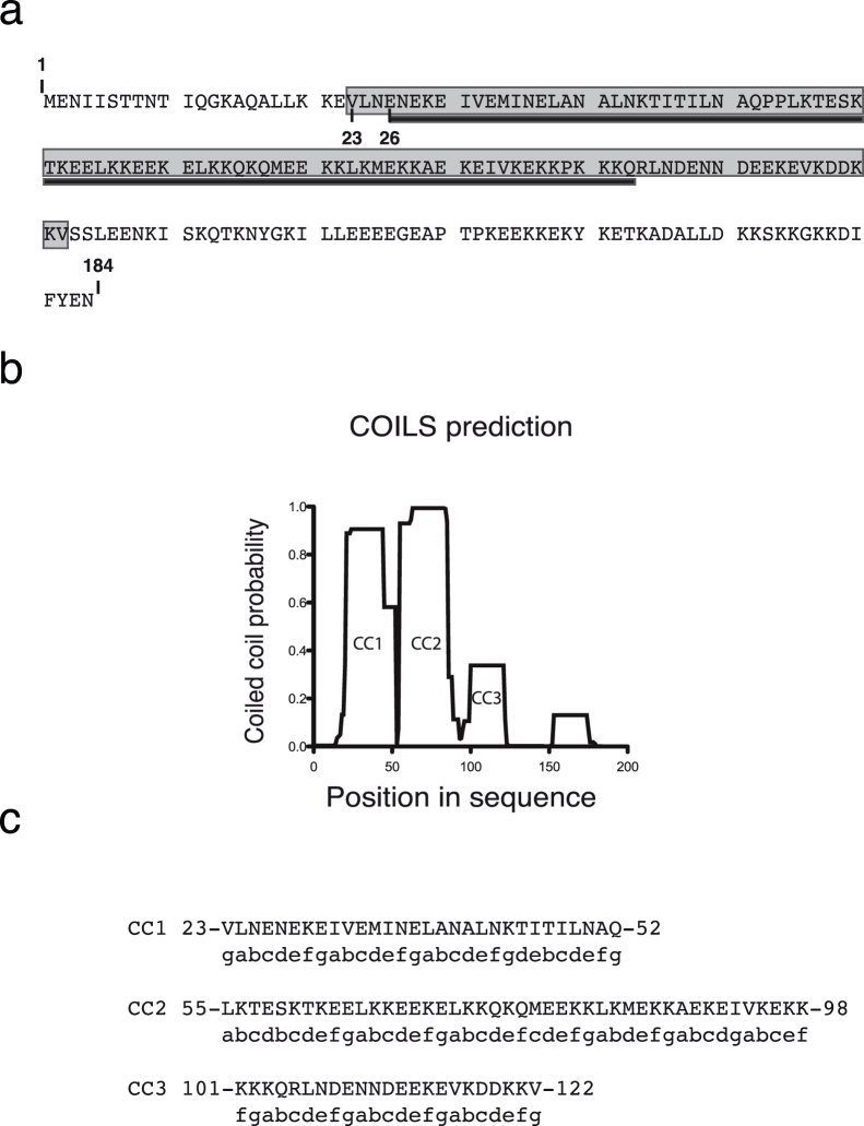
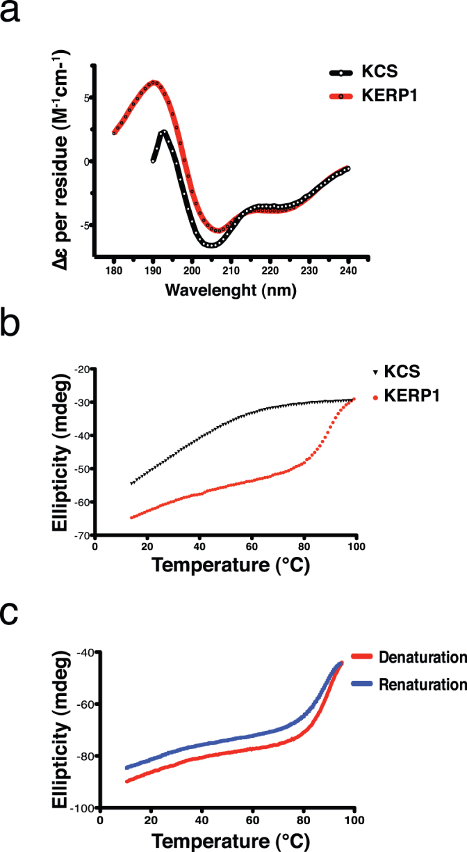
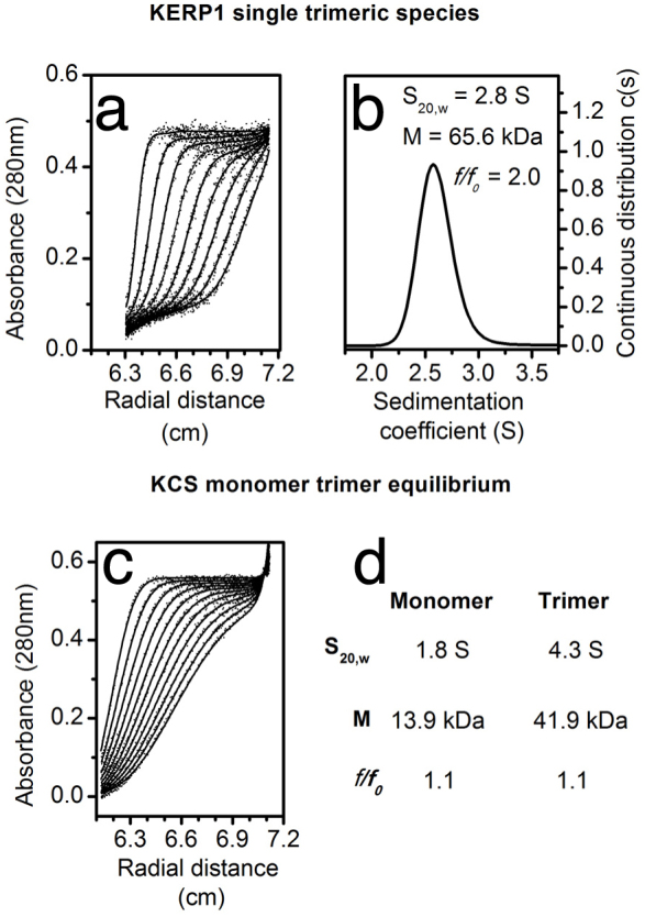
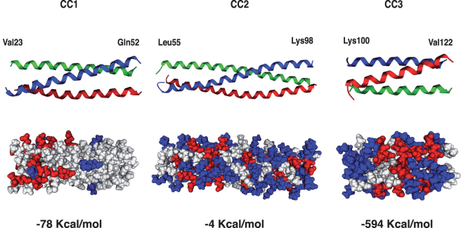
The lysine and glutamic acid rich protein KERP1 is a unique surface adhesion factor associated with virulence in the human pathogen Entamoeba histolytica. Both the function and structure of this protein remain unknown to this date. Here, we used circular dichroism, analytical ultracentrifugation and bioinformatics modeling to characterize the structure of KERP1. Our findings revealed that it is an α-helical rich protein organized as a trimer, endowed with a very high thermal stability (Tm = 89.6°C). Bioinformatics sequence analyses and 3D-structural modeling indicates that KERP1 central segments could account for protein trimerization. Relevantly, expressing the central region of KERP1 in living parasites, impair their capacity to adhere to human cells. Our observations suggest a link between the inhibitory effect of the isolated central region and the structural features of KERP1.


Figures

Figure 1
Figure 1. KERP1 protein domains predicted by bioinformatics analysis.
(a). Analysis of KERP1 primary structure using the COILS and Pfam servers. The coiled-coil domain (KCS) is highlighted in grey (from amino acid 23 to 122) and the Universal Stress Protein (Usp) domain (amino acid 26 to 103) is underlined in black. (b). Graphical representation of the COILS server prediction output using the complete amino acid sequence of KERP1 (Accession number EHI_098210). (c). Coiled-coil domain segments of KERP1 CC1 (residues 23 to 52), CC2 (residues 55 to 98) and CC3 (residues 101 to 122) are represented using the heptad repeat (a-b-c-d-e-f-g)n assignation from COILS prediction.
Figure 2
Figure 2. KCS is expressed in E. histolytica transfectants and localizes in the same cellular compartments as KERP1.
Biochemical detection of KERP1 and KCS in transfected trophozoites TetOCat and TetOCat-KCS. (a). Immunoblotting of parasite protein extracts at 48 hours of tetracycline induction (+) or without (−). Detection of endogenous actin (49 kDa; mAb anti-actin C4) was used as protein extract loading control. Detection of KERP1 (25 kDa; mAb anti-KERP1 C2-7) in all strains was observed, but detection of HSV-KCS (15 kDa; mAb anti-HSVtag) was only remarked in TetOCat-KCS (+). Immunoblotting was performed using simultaneously all the primary antibodies. (b). Endogenous KERP1 colocalizes with KCS in live parasites. Cellular detection of KERP1 and KCS in transfected trophozoites TetOCat and TetOCat-KCS after 48 hours of tetracycline induction. Micrographs showing immunofluorescence images acquired by confocal microscopy. KERP1 is detected by a specific monoclonal antibody (green; mAb C2-7), KCS by the HSV-tag (red; mAb anti-HSVtag) and nuclei are labeled with DAPI (blue). KERP1 and KCS share the same cellular compartment as observed by the concentrated patches (yellow) at the trophozoite plasma membrane and internal vesicles. Focal planes from a Z-stack were selected. Scale bar 10 µm.
Figure 3
Figure 3. Expression of KERP1 coiled-coil domains in E. histolytica reduces the capacity to adhere to human cells without affecting cytotoxicity.
Functional assays of adherence and cytotoxicity using E. histolytica transfectants TetOCat and TeTOCat-KCS with (+) or without (−) induction by tetracycline, interacting with human cells LSEC or Caco2. Assays were performed in a ratio of 1:5 amoebas to human Caco2 cells and 1:10 to human LSEC. (a). Percentage values of adherence to LSEC (p value: 0.004). (b). Percentage values of adherence to Caco2 cells (p value: 0.002). A statistically significant reduction of 60% in adherence is observed for trophozoites expressing KCS (TetOCat-KCS+) compared to the control (TetOCat), independently of the human cell type. Cytotoxicity assays were performed using the same ratio as before with a control sample without parasites, indicating the 100% rate of survival of the human cells during the experiment. (c). Percentage values of LSEC survival (p value *: 0.02; **: 0.006). (d). Percentage values of Caco2 cell survival. No difference in the percentage of survival between the different human cell lines was observed upon comparison with the control.
Figure 4
Figure 4. KERP1 is an α-helical protein highly stable to thermal denaturation.
(a). KERP1 and KCS secondary structure analysis determined by following circular dichroism signal in the far–UV region. KERP1 shows two negative peaks minima at 222 nm and 208 nm that highlight the content of α-helices in the protein (red) contrary to KCS with two negative peaks minima at 222 nm and 205 nm that highlight the content of unstructured regions in the protein (black). (b). Thermal denaturation of KERP1 was observed by the loss in ellipticity at 222 nm when increasing temperature (10° to 100°C). The thermal melting point was calculated using a two-state cooperative transition obtaining a value of 89.6°C for KERP1 (red) and 60°C for KCS (black). (c). Melting curves for KERP1, measured by monitoring the absorbance at 222 nm against increasing (denaturation) and decreasing temperatures (renaturation).
Figure 5
Figure 5. Analytical ultracentrifugation characterization of KERP1 and KCS.
Sedimentation velocity analysis of KERP1 (a and b) and KCS (c and d) at 20 µM. (a) Radial distribution of optical density measurements at 280 nm of KERP1: experimental data (dot), and fitted line (RMSD <0.006) to the continuous distribution model. (b) Sedimentation coefficient distribution c(s) showing a single peak compatible with a trimeric form of KERP1 with its hydrodynamic properties. (c) Radial distribution of optical density measurements at 280 nm of KCS experimental data (dot), and fitted line (RMSD <0.005) to the self-association monomer/trimer model. (d) Hydrodynamic properties of KCS monomer and trimer.
Figure 6
Figure 6. Three-dimensional structure prediction of KERP1 trimeric coiled-coil regions.
Three-dimensional modeling of the CC regions predicted for KERP1 according to COILS results. CC regions of KERP1 correspond to the KCS protein designated as, CC1 (Valine23 to Glutamine52), CC2 (Leucine55 to Lysine98) and CC3 (Lysine100 to Valine122), folded as elongated trimer. Modeling was performed using chain A of PDB file ID 1WT6 as template with its distinctive trimeric coiled-coil organization. Each of the three alpha helical ribbon chains is colored differently in order to differentiate them. Under the ribbons CPK model highlight the presence of positively charged residues (blue) and negatively charged residues (red). The most stable coiled coil trimer corresponds to CC3 (residues Lys-100 to Val-122) according to an electrostatic analysis on each of the CC predicted regions and the calculated values of free energy present for each, represented in the bottom of the CPK models.

Similar articles

Cited by

References

    1. Stanley S. L. Jr Amoebiasis. Lancet 361, 1025–1034 (2003). - PubMed
    1. Seigneur M., Mounier J., Prevost M. C. & Guillen N. A lysine- and glutamic acid-rich protein, KERP1, from Entamoeba histolytica binds to human enterocytes. Cell Microbiol 7, 569–579 (2005). - PubMed
    1. Santi-Rocca J. et al. The lysine- and glutamic acid-rich protein KERP1 plays a role in Entamoeba histolytica liver abscess pathogenesis. Cell Microbiol 10, 202–217 (2008). - PubMed
    1. Burkhard P., Stetefeld J. & Strelkov S. V. Coiled coils: a highly versatile protein folding motif. Trends Cell Biol 11, 82–88 (2001). - PubMed
    1. Parry D. A., Fraser R. D. & Squire J. M. Fifty years of coiled-coils and alpha-helical bundles: a close relationship between sequence and structure. J Struct Biol 163, 258–269 (2008). - PubMed

Publication types

MeSH terms

Substances

LinkOut - more resources