. 2013 Mar 12;52(10):1725-36.
doi: 10.1021/bi3012077. Epub 2013 Mar 1.

Type II kinase inhibitors show an unexpected inhibition mode against Parkinson's disease-linked LRRK2 mutant G2019S

Affiliations

Type II kinase inhibitors show an unexpected inhibition mode against Parkinson's disease-linked LRRK2 mutant G2019S

Min Liu et al. Biochemistry. .

Abstract

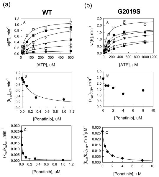
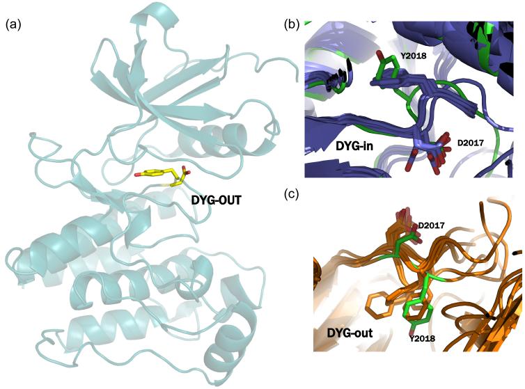
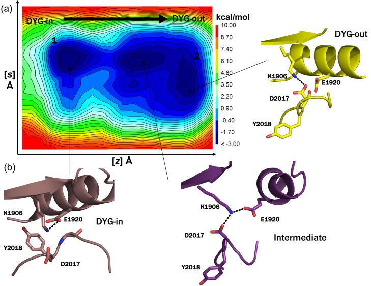
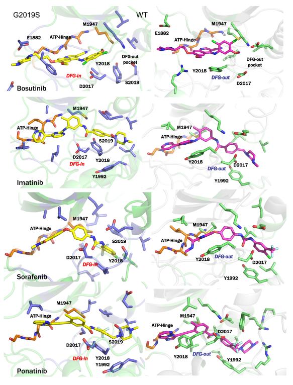
A number of well-known type II inhibitors (ATP-noncompetitive) that bind kinases in their DFG-out conformation were tested against wild-type LRRK2 and the most common Parkinson's disease-linked mutation, G2019S. We found that traditional type II inhibitors exhibit surprising variability in their inhibition mechanism between the wild type (WT) and the G2019S mutant of LRRK2. The type II kinase inhibitors were found to work in an ATP-competitive fashion against the G2019S mutant, whereas they appear to follow the expected noncompetitive mechanism against WT. Because the G2019S mutation lies in the DXG motif (DYG in LRRK2 but DFG in most other kinases) of the activation loop, we explored the structural consequence of the mutation on loop dynamics using an enhanced sampling method called metadynamics. The simulations suggest that the G2019S mutation stabilizes the DYG-in state of LRRK2 through a series of hydrogen bonds, leading to an increase in the conformational barrier between the active and inactive forms of the enzyme and a relative stabilization of the active form. The conformational bias toward the active form of LRRK2 mutants has two primary consequences. (1) The mutant enzyme becomes hyperactive, a known contributor to the Parkinsonian phenotype, as a consequence of being "locked" into the activated state, and (2) the mutation creates an unusual allosteric pocket that can bind type II inhibitors but in an ATP-competitive fashion. Our results suggest that developing type II inhibitors, which are generally considered superior to type I inhibitors because of desirable selectivity profiles, might be especially challenging for the G2019S LRRK2 mutant.


Figures

Figure 1
Figure 1
(a) Inhibition study of wt LRRK2 catalyzed phosphorylation of LRRKtide by Ponatinib. A: Plot of initial velocities vs [ATP] at [Ponatinib] = 1 (●), 0.3 (엯), 0.1 (▾), 0.04 (▿), 0.1 (∎), and 0 μM (□) all at a fixed LRRKtide concentration of 50 μM. B & C: Ponatinib concentration dependencies of (kcat)ATP and (kcat/Km)ATP apparent values derived from analysis of the data of panel A. (b) Inhibition study of the G2019S mutant catalyzed phosphorylation of LRRKtide by Ponatinib. A: Plot of initial velocities vs [ATP] at [Ponatinib] = 8 (●), 4 (엯), 2 (▾), 1 (▿), 0.5 (∎), and 0 μM (□) all at a fixed LRRKtide concentration of 50 μM. B & C: Ponatinib concentration dependencies of (kcat)ATP and (kcat/Km)ATP apparent values derived from analysis of the data of panel A.
Figure 2
Figure 2
Temperature dependencies for WT LRRK2 and mutant-catalyzed LRRKtide and LRRKtideS phosphorylation. The Eyring plots show kcat dependence of temperature for WT LRRK2 (a), the mutant G2019S (b). The kcat was the average of four independent measurements. The data from these plots are summarized in table 1.
Figure 3
Figure 3
(a) Homology of model of LRRK2 with the activation loop in the DYG-out conformation generated using Prime. (b) superposition of the DYG-in model of LRRK2 with x-ray structures of cABL, cKIT, Aurora, B-raf, EPHA3, SRC, LCK and MK14 in their DFG-in conformation. The overall RMSD between any two pairs of structures is < 1.9Å and the DYG-in of LRRK2 (shown in green) model is conformationally very similar to these x-ray structures. (c) superposition of the DYG-out model of LRRK2 (shown in green) generated using Prime with x-ray structures of cABL, cKIT, Aurora, B-raf, EPHA3, SRC, LCK and MK14 in their DFG-out conformation. The overall RMSD between any two pairs of structures is < 2.0Å and the DYG-out of LRRK2 model is conformationally very similar to these x-ray structures.
Figure 4
Figure 4
(a) Representative free energy surface (FES) generated from 250 ns metadynamics simulation of WT LRRK2 generated by following the opening and closing of the enzyme involving the ATP binding β-sheet domain and C-helix as the first collective variable (z) and the motion of the center of mass of the activation loop as the second collective variable (s) as the kinase switches between active and inactive form. The FES shows that opening and closing motion of the enzyme occurs unhindered and the activation loop can switch between the active and inactive form easily (a contagious low energy path connects the two conformations forms). (b) Snapshots of structures extracted from local clustering of conformations within the simulation corresponding to the three local minima observed during the course of the simulation are shown here. The states 1 and 2 correspond to the active (DYG-in) and inactive (DYG-out) states of the enzyme. An intermediate metastable transition is observed, where K1906 makes a shared hydrogen bond with both E1920 (C-helix) and D2017 (DYG-motif).
Figure 5
Figure 5
(a) Representative free energy surface (FES) generated from a 250 ns metadynamic simulation run of G2019S mutant generated by following the opening and closing of the enzyme involving the ATP binding β-sheet domain and C-helix as the first collective variable (z) and the motion of the center of mass of the activation loop as the second collective variable (s) as the kinase switches between active and inactive form. The simulations were carried out under identical conditions as that of the WT LRRK2 described above. The FES plot shows that there is a high energy barrier that separates the active and inactive form of the kinases indicating that higher amount of energy is required for the mutant to switch between active and inactive conformations relative to WT. However, independently both the active and inactive form of the enzyme can switch between open and closed forms. (b) Snapshots of structures extracted from local clustering of conformations within the simulation corresponding to the three local minima are shown here. In state 1, the G2019S mutation results in a side-chain of E1920 and S2019. This offers additional stability to this state. In state 2, the side-chain of S2019 makes a hydrogen bond with the backbone of D2017 and appears to be a metastable state. A transient stable intermediate state 3 is observed in the simulation where the side chain of S2019 makes a hydrogen bond with backbone of Y2018.
Figure 6
Figure 6
(left panels) Structural representations of induced fit docked conformations for the known DFG-out inhibitors bosutinib, imatinib, sorafenib and ponatinib on the DFG-in conformation of G2019S mutant of LRRK2. (right panels) Structural representations of induced fit docked conformations for the known DFG-out inhibitors bosutinib, imatinib, sorafenib and ponatinib on the DFG-out conformation of WT LRRK2. In this figure, only few residues are labeled to preserve clarity. Figure S5 in Supporting Information accompanies this figure showing all protein inhibitor interactions.

References

    1. Bosco DA, LaVoie MJ, Petsko GA, Ringe D. Proteostasis and movement disorders: Parkinson’s disease and amyotrophic lateral sclerosis. Cold Spring Harb Perspect Biol. 2011;3:a007500. - PMC - PubMed
    1. Martin I, Dawson VL, Dawson TM. Recent Advances in the Genetics of Parkinson’s Disease. Annu Rev Genomics Hum Genet. 2010 - PMC - PubMed
    1. Hardy J, Cookson MR, Singleton A. Genes and parkinsonism. Lancet Neurol. 2003;2:221–228. - PubMed
    1. Cookson MR. Pathways to Parkinsonism. Neuron. 2003;37:7–10. - PubMed
    1. Benamer HT. The ancestry of LRRK2 Gly2019Ser parkinsonism. Lancet Neurol. 2008;7:769–770. author reply 770-761. - PubMed

MeSH terms

Substances