. 2013 Mar;171(3):283-97.
doi: 10.1111/cei.12015.

Immunotherapeutic effects of recombinant adenovirus encoding granulocyte-macrophage colony-stimulating factor in experimental pulmonary tuberculosis

Affiliations

Immunotherapeutic effects of recombinant adenovirus encoding granulocyte-macrophage colony-stimulating factor in experimental pulmonary tuberculosis

A Francisco-Cruz et al. Clin Exp Immunol. 2013 Mar.

Abstract

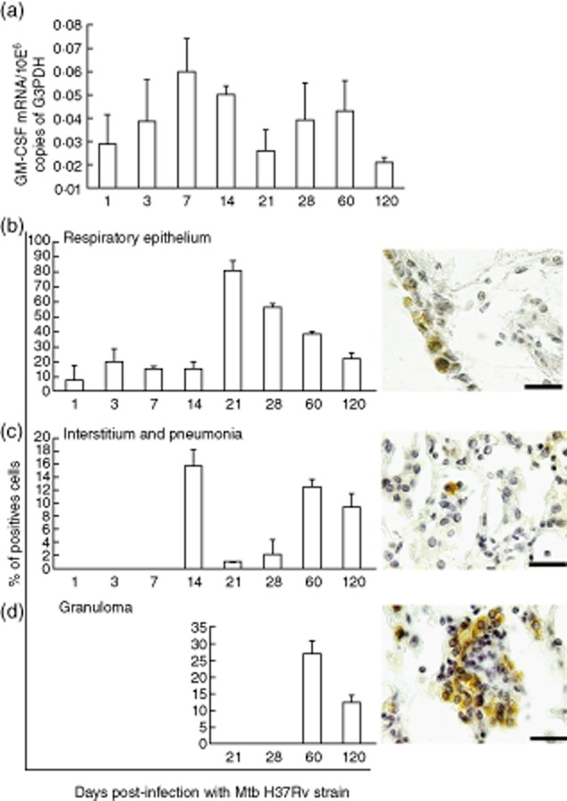
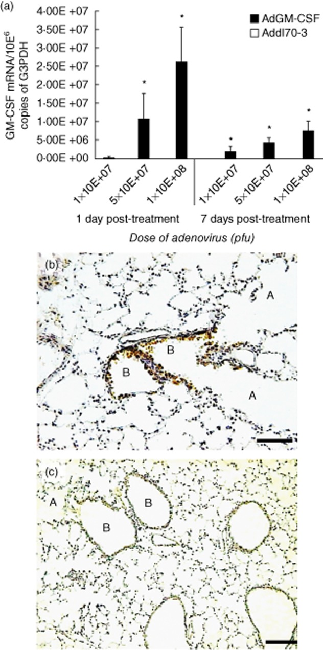
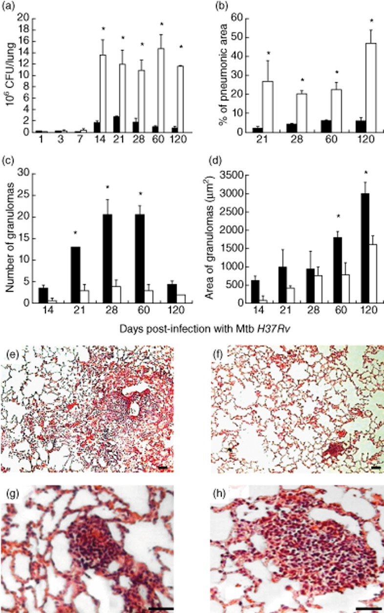
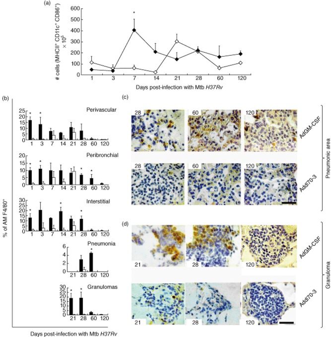
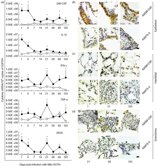
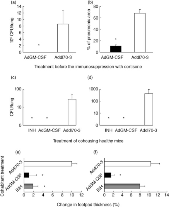
BALB/c mice with pulmonary tuberculosis (TB) develop a T helper cell type 1 that temporarily controls bacterial growth. Bacterial proliferation increases, accompanied by decreasing expression of interferon (IFN)-γ, tumour necrosis factor (TNF)-α and inducible nitric oxide synthase (iNOS). Activation of dendritic cells (DCs) is delayed. Intratracheal administration of only one dose of recombinant adenoviruses encoding granulocyte-macrophage colony-stimulating factor (AdGM-CSF) 1 day before Mycobacterium tuberculosis (Mtb) infection produced a significant decrease of pulmonary bacterial loads, higher activated DCs and increased expression of TNF-α, IFN-γ and iNOS. When AdGM-CSF was given in female mice B6D2F1 (C57BL/6J X DBA/2J) infected with a low Mtb dose to induce chronic infection similar to latent infection and corticosterone was used to induce reactivation, a very low bacilli burden in lungs was detected, and the same effect was observed in healthy mice co-housed with mice infected with mild and highly virulent bacteria in a model of transmissibility. Thus, GM-CSF is a significant cytokine in the immune protection against Mtb and gene therapy with AdGM-CSF increased protective immunity when administered in a single dose 1 day before Mtb infection in a model of progressive disease, and when used to prevent reactivation of latent infection or transmission.


Figures

Figure 1
Figure 1
Kinetics of granulocyte–macrophage colony-stimulating factor (GM-CSF) during progressive pulmonary tuberculosis (TB). (a) Five lungs from the same number of Mycobacterium tuberculosis (Mtb)-infected mice at each indicated time-point were used to isolate total RNA and determine the gene expression of GM-CSF by quantitative reverse transcription–polymerase chain reaction (RT–PCR). (b–d) GM-CSF protein expression was detected by immunohistochemistry and the percentage of positive cells was determined in the indicated lung compartments by automated morphometry; representative histological figures of each compartment are to the right of the morphometry graphs (scale bar represents 20 μm). All values are means ± standard deviation (s.d.) from two independent experiments with five mice per group (408 × 552 mm; 150 × 150 DPI).
Figure 2
Figure 2
Granulocyte–macrophage colony-stimulating factor (GM-CSF) expression after intratracheal (i.t.) administration of different doses of adenoviruses encoding GM-CSF (AdGM-CSF). (a) Groups of healthy mice were treated with the indicated dose of AdGM-CSF (black bars) or the control adenoviruses Addl70-3 (white bars, undetectable levels), and euthanized after 1 and 7 days; the lungs were used to determine the expression of GM-CSF reverse transcription–polymerase chain reaction (RT–PCR), values are means ± standard deviation (s.d.) from two independent experiments with five mice per group; *P < 0·05. (b) GM-CSF protein expression was detected by immunohistochemistry; the mouse lung after 1 day of 1 × 10⁸ plaque-forming units (pfu) i.t. administration shows strong immunostaining in the airways epithelium in bronchioles (B) and negative immunostaining in alveolar walls (A). (c) In contrast, the mouse lung treated with the same dose of control adenovirus Addl70-3 does not show immunostaining (scale bar represents 60 μm) (207 × 386 mm; 300 × 300 DPI).
Figure 3
Figure 3
Effect of a single administration of adenoviruses encoding granulocyte–macrophage colony-stimulating factor (AdGM-CSF) 1 day before infection in the murine model of progressive pulmonary tuberculosis (TB). (a) Groups of mice were treated with AdGM-CSF (black bars) or control virus Addl70-3 (white bars) 1 day before intratracheal (i.t.) infection with a high dose of Mycobacterium tuberculosis (Mtb) H37Rv; five mice were killed at each day indicated and the lungs were used to determine bacterial loads by colony-forming units (CFU). A significant CFU decrease was produced by AdGM-CSF administration. (b) The morphometry study showed a significantly lower percentage of lung surface affected by pneumonia in animals treated with AdGM-CSF. (c) The number of granulomas at days 21, 28 and 60 was significantly higher in the AdGM-CSF-treated group, as well as the granuloma size at days 60 and 120 (d). Representative histopathology of the lung from treated mice after 120 days of infection with Mtb, (e) the mouse lung treated with Addl70-3 shows extensive areas of pneumonia. (f) In contrast, mouse treated with AdGM-CSF shows scarce inflammation. (g) Small granulomas are seen in mice treated with Addl70-3. (h) In comparison, bigger granulomas are formed in mouse treated with AdGM-CSF; scale bars represent 60 μm (e) and (f), and 20 μm (g) and (h). All values of bacilli loads and histomorphometry are means ± standard deviation (s.d.) of two independent experiments with five mice per group; *P < 0·05 (466 × 771 mm; 150 × 150 DPI).
Figure 4
Figure 4
Effect of a single administration of adenoviruses encoding granulocyte–macrophage colony-stimulating factor (AdGM-CSF) on the production of activated dendritic cells and macrophages in the murine model of progressive pulmonary tuberculosis (TB). (a) Groups of mice were treated with AdGM-CSF (black symbols) or Addl70-3 (white symbols) 1 day before infection with Mycobacterium tuberculosis (Mtb) H37Rv; lungs were used to obtain cell suspensions and the number of activated dendritic cells was determined by flow cytometry using antibodies against MHC II-fluorescein isothiocyanate (FITC), CD11c-allophycocyanin (APC) and CD86-phycoerythrin (PE). For each lung sample 1 × 10⁵ events were acquired and the population of cells positive for FITC (FL1) and APC (FL4) (i.e. MHC II+ CD11c+ cells) were selected for obtaining the histogram for the number of positive events for PE (FL2) (i.e. MHC II+ CD11c+ CD86+ cells). The treatment with AdGM-CSF induced more rapid and higher numbers of activated dendritic cells. (b) Lung sections from treated animals with adenoviruses were used to detect the marker of activated macrophages F4/80 by immunohistochemistry, and the percentage of immunostained cells were determined in the indicated lung compartments and lesions. Results are expressed as means ± standard deviation (s.d.) from five mice per time-point and per group in two independent experiments. Representative immunohistochemistry detection of F4/80+ cells in the lungs of animals treated with AdGM-CSF or Add170-3 at the indicated time-point (left corner) in pneumonic areas (c) or granulomas (d). There are more F4/80+ cells in animals treated with AdGM-CSF; scale bar represents 20 μm (518 × 553 mm; 200 × 200 DPI).
Figure 5
Figure 5
Effect of a single administration of adenoviruses encoding granulocyte–macrophage colony-stimulating factor (AdGM-CSF) on the cytokines and inducible nitric oxide synthase (iNOS) gene transcription in the murine model of progressive pulmonary tuberculosis (TB). (a) Groups of mice infected by the intratracheal (i.t.) route with a high dose of Mycobacterium tuberculosis (Mtb) to induce progressive TB were treated 1 day before with AdGM-CSF (black symbols) or Addl70-3 (white symbols), and their lungs were used to isolate total RNA and quantify the expression of the indicated cytokines by real-time reverse transcription–polymerase chain reaction (RT–PCR). Animals treated with AdGM-CSF showed a higher expression of cytokines and iNOS. Results are expressed as means ± standard deviation (s.d.) of two independent experiments with five mice per group; *P < 0·05. Representative immunohistochemistry of GM-CSF cellular detection in treated animals with AdGM-CSF or Add170-3 at different time-points in bronchial epithelium (b), alveolar–capillary interstitium (c) and granulomas (d); more immunostained cells are observed in animals treated with AdGM-CSF, the strongest being labelled the airways epithelium; scale bar represents 20 μm (439 × 475 mm; 200 × 200 DPI).
Figure 6
Figure 6
Adenovirus encoding granulocyte–macrophage colony-stimulating factor (AdGM-CSF) treatment prevents reactivation of chronic tuberculosis (TB) similar to latent infection following immunosuppression and prevention of transmission in co-housed healthy mice. (a) Mice were infected with low-dose Mycobacterium tuberculosis (Mtb) H37Rv to induce chronic infection similar to latent infection, and after 7 months animals were treated with AdGM-CSF (black bars) or Addl70-3 (white bars); after 1 month mice were treated with cortisone for 1 month to induce disease reactivation. Animals treated with AdGM-CSF showed lower pulmonary bacilli loads and (b) lesser tissue damage (pneumonia) than mice treated with Addl70-3. (c) With regard to evaluation of the effect of AdGM-CSF treatment in the prevention of transmission, groups of mice were infected with mild virulent Mtb strain H37Rv or (d) with the highly virulent Beijing strain 9001000 and co-housed with healthy mice treated with isoniazid (INH; hatched bar), AdGM-CSF (black bar) or Addl70-3(white bar) for 2 months; the lungs were used for bacilli burden determination by quantification of colony-forming units (CFUs). In comparison with animals treated with Addl70-3, the lungs of animals treated with INH or AdGM-CSF do not show bacilli growth. (e) Significantly lower delayed-type hypersensitivity (DTH) was measured in animals treated with INH or AdGM-CSF than mice treated with Addl70-3 that were co-housed with mice infected with Mtb strain H37Rv. (f) In contrast, there was no DTH difference in animals treated with INH or Addl70-3 when co-housed with mice infected with highly virulent Beijing strain, while animals treated with AdGM-CSF showed significantly lower DTH. All values are means ± standard deviation (s.d.) from two independent experiments with five mice per group;*P < 0·05 (561 × 632 mm; 200 × 200 DPI).

Similar articles

Cited by

References

    1. Geneva, Switzerland: World Health Organization; 2009. Global tuberculosis control: epidemiology, strategy, financing. WHO report 2009.
    1. Parrish NM, Dick JD, Bishai WR. Mechanisms of latency in Mycobacterium tuberculosis. Trends Microbiol. 1998;6:107–112. - PubMed
    1. Tufariello J, Chan J, Flynn J. Latent tuberculosis: mechanisms of host and bacillus that contribute to persistent infection. Lancet Infect Dis. 2003;3:578–590. - PubMed
    1. Kaufmann SH. How can immunology contribute to the control of tuberculosis? Nat Rev Immunol. 2001;1:20–30. - PubMed
    1. Nakaoka H, Lawson L, Squire SB. Risk for tuberculosis among children. Emerg Infect Dis. 2006;12:1383–1388. et al. - PMC - PubMed

Publication types

Substances