

doi: 10.1021/nn305030g.
Epub 2013 Feb 4.
Nuclease-resistant DNA via high-density packing in polymeric micellar nanoparticle coronas
Affiliations
- PMID: 23379679
- PMCID: PMC3608424
- DOI: 10.1021/nn305030g
Item in Clipboard
Nuclease-resistant DNA via high-density packing in polymeric micellar nanoparticle coronas
ACS Nano.
.
Abstract
Herein, we describe a polymeric micellar nanoparticle capable of rendering nucleic acids resistant to nuclease digestion. This approach relies on utilizing DNA as the polar headgroup of a DNA-polymer amphiphile in order to assemble well-defined, discrete nanoparticles. Dense packing of DNA in the micelle corona allows for hybridization of complementary oligonucleotides while prohibiting enzymatic degradation. We demonstrate the preparation, purification, and characterization of the nanoparticles, then describe their resistance to treatment with endo- and exonucleases including snake-venom phosphodiesterase (SVP), a common, general DNA digestion enzyme.
Figures
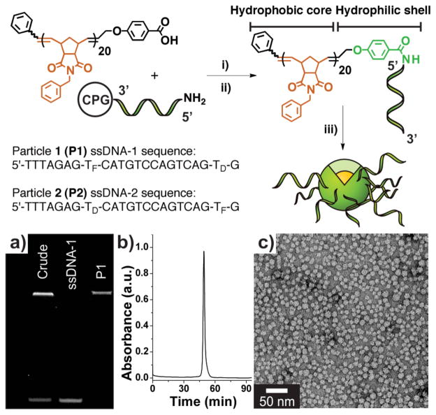
Preparation of DNA-polymer amphiphiles (DPAs) and assembly of micelles. Synthesis: i) A hydrophobic polymer, terminally modified with a carboxylic acid moiety was mixed with a coupling agent and reacted with a 5′-amino modified oligonucleotide on solid support (controlled pore glass, CPG). ii) Deprotection and cleavage of the resulting DNA-polymer conjugate from solid support. iii) Dialysis of cleaved DPA into deionized water to form a mixture of micelles and free, non-conjugated nucleic acid. TF and TD correspond to fluorescein- and DABCYL-modified thymidine phosphoramidites. a) PAGE analysis. Lane 1: Crude material post-micelle (P1) formation showing conjugate (top band) and free ssD-NA (lower band). Lane 2: HPLC purified sample of ssDNA-1. Lane 3: Purified P1, isolated via size-exclusion chromatrography (SEC). b) SEC trace of purified P1 (λabs = 260 nm). c) Transmission electron micrograph of P1. See Supporting Information Figure S7 for P2 data.
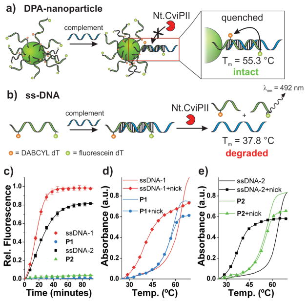
Endonuclease resistance of DPA nanoparticles. a) Scheme depicting DPA-nanoparticle (P2) resistance to nicking endonuclease (Nt.CviPII) and consequently intact, quenched duplex DNA at the particle surface. b) Scheme depicting dsDNA degradation by Nt.CviPII and consequently a decrease in duplex melting temperature and increase in fluorescein fluorescence. c) Nt.CviPII activity over time, monitored via fluorescein fluorescence dequenching (λex = 485 nm, λem = 535 nm). d) Thermal denaturation analysis with and without Nt.CviPII treatment for P1 and ssDNA-1. λabs = 260 nm. Sample subjected to enzyme for 100 minutes at 37 °C. ssDNA-1 + Complement: Tm = 63.9 °C; ssDNA-1 + Nt.CviPII + Complement: Tm = 37.8 °C; P1 + Complement: Tm = 58.8 °C; P1 + Nt.CviPII + Complement: Tm = 58.3 °C). e) Thermal denaturation analysis with and without Nt.CviPII treatment for P2 and ssDNA-2. λabs = 260 nm. Sample subjected to enzyme for 100 minutes at 37 °C. ssDNA-2 + Complement: Tm = 63.9 °C; ssDNA-2 + Nt.CviPII + Complement: Tm = 37.8 °C; P2 + Complement: Tm = 56.9 °C; P2 + Nt.CviPII + Complement: Tm = 55.3 °C). See Supporting Information Figure S12 for derivative plots of melting temperatures. Complement: 5′-TATTATATCTTTAGACACTGA CTGGACATGACTCT-3′
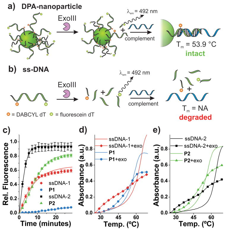
Exonuclease resistance of DPA nanoparticles. a) Scheme depicting DPA-nanoparticle resistance to Exonuclease III and consequently, intact DNA at the particle surface available for hybridization with complementary ssDNA. b) Scheme depicting ssDNA being degraded by ExoIII and consequently, no intact DNA available for hybridization with complementary ssDNA. c) Exonuclease III activity over time monitored by fluorescein fluorescence dequenching (λex = 485 nm, λem = 535 nm). d) Thermal denaturation analysis with and without Exo III treatment for P1 and ssDNA-1. λabs = 260 nm. Samples subjected to enzyme for 60 minutes at 37 °C. P1 + Complement: Tm = 58.8 °C; P1 + Exo III + Complement: Tm = 55.8 °C). e) Thermal denaturation analysis with and without Exo III treatment for P2 and ssDNA-2. λabs = 260 nm. Samples subjected to enzyme for 60 minutes at 37 °C. P2 + Complement: Tm = 56.9 °C; P2 + Exo III + Complement: Tm = 53.9 °C). See Supporting Information Figure S13 for derivative plots of melting temperatures. Complement: 5′-TATTATATCTTTAGACAC TGACTGGACATGACTCT-3′
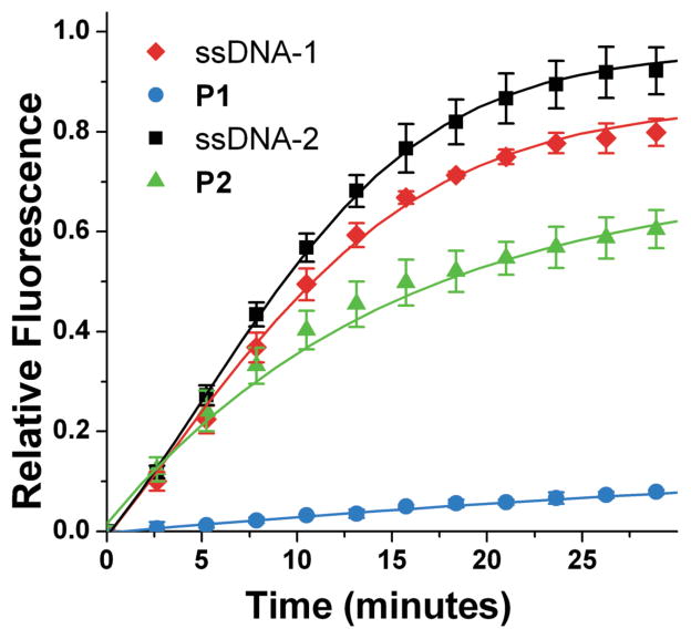
SVP activity over time monitored by fluorescein fluorescence dequenching (λex = 485 nm, λem = 535 nm).
References
-
- Winfree E, Liu F, Wenzler LA, Seeman NC. Design and Self-Assembly of Two-Dimensional DNA Crystals. Nature. 1998;394:539–544. - PubMed
-
- Akhtar S, Hughes MD, Khan A, Bibby M, Hussain M, Nawaz Q, Double J, Sayyed P. The Delivery of Antisense Therapeutics. Adv Drug Deliv Rev. 2000;44:3–21. - PubMed
-
- Thomas M, Klibanov AM. Non-Viral Gene Therapy: Polycation-Mediated DNA Delivery. App Microbiol Biotechnol. 2003;62:27–34. - PubMed
-
- Davis ME. Non-Viral Gene Delivery Systems. Curr Opinion Biotechnol. 2002;13:128–131. - PubMed
-
- Leumann CJ. DNA Analogues: From Supramolecular Principles to Biological Properties. Bioorg Med Chem. 2002;10:841–854. - PubMed
Publication types
MeSH terms
Substances
Grants and funding
LinkOut - more resources
Full Text Sources
Other Literature Sources
