

IL-22 is related to development of human colon cancer by activation of STAT3
- PMID: 23379788
- PMCID: PMC3607898
- DOI: 10.1186/1471-2407-13-59
IL-22 is related to development of human colon cancer by activation of STAT3
Abstract
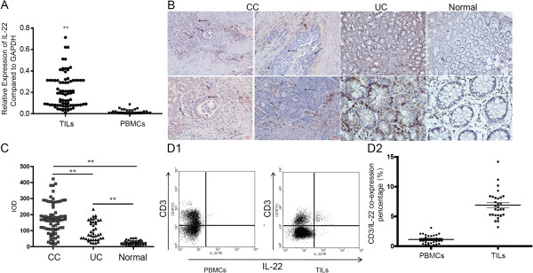
Background: It has been previously reported that IL-22, one of the cytokines secreted by Th17 cells, demonstrates both a protective and inflammatory promotion effect in inflammatory bowel disease (IBD) through STAT3 signaling activation. We sought to investigate the role of IL-22 expression in colon cancer (CC).
Methods: The expression of IL-22 and related molecules were detected in human CC, the detail function and mechanism of IL-22 were investigated by in vivo and in vitro model.
Results: Our results demonstrated significant upregulation of IL-22 in human CC tumor infiltrated leukocytes (TILs) compared to peripheral lymphocytes. Moreover, our findings demonstrated that IL-22 expression was significantly higher in ulcerative colitis (UC) tissues versus normal colon tissues. Both IL-22 receptor α1 (IL-22RA1) and IL-23 were highly expressed in CC and UC tissues compared to normal controls. TILs exhibiting various IL-22 expression levels isolated from CC patients were demonstrated to enhance tumor growth and metastasis co-transplanted with Hct-116 cells underwent subcutaneous transplantation in mice model. Tumor growth and metastasis was promoted by STAT3 phosphorylation and upregulation of its downstream genes such as Bcl-xl, CyclinD1, and VEGF. In vitro studies confirmed the anti-apoptotic and pro-proliferation effect of IL-22 according to the BrdU cooperation assay and peroxide induced apoptosis analysis with or without the presence of IL-22.
Conclusion: In this study we demonstrated that excessive IL-22 in the CC and UC microenvironment leads to tumor growth, inhibition of apoptosis, and promotion of metastasis depend on STAT3 activation.
Figures


Similar articles
-
Interleukin-22 promotes human hepatocellular carcinoma by activation of STAT3.Hepatology. 2011 Sep 2;54(3):900-9. doi: 10.1002/hep.24486. Epub 2011 Aug 8. Hepatology. 2011. PMID: 21674558
-
Expression of interleukin-22/STAT3 signaling pathway in ulcerative colitis and related carcinogenesis.World J Gastroenterol. 2013 May 7;19(17):2638-49. doi: 10.3748/wjg.v19.i17.2638. World J Gastroenterol. 2013. PMID: 23674871 Free PMC article.
-
Th22 cell accumulation is associated with colorectal cancer development.World J Gastroenterol. 2015 Apr 14;21(14):4216-24. doi: 10.3748/wjg.v21.i14.4216. World J Gastroenterol. 2015. PMID: 25892871 Free PMC article.
-
High suppressor of cytokine signaling-3 expression impairs STAT3-dependent protective effects of interleukin-22 in ulcerative colitis in remission.Inflamm Bowel Dis. 2015 Feb;21(2):241-50. doi: 10.1097/MIB.0000000000000267. Inflamm Bowel Dis. 2015. PMID: 25545374
-
Th17 cells inhibit CD8+ T cell migration by systematically downregulating CXCR3 expression via IL-17A/STAT3 in advanced-stage colorectal cancer patients.J Hematol Oncol. 2020 Jun 5;13(1):68. doi: 10.1186/s13045-020-00897-z. J Hematol Oncol. 2020. PMID: 32503584 Free PMC article.
Cited by
-
IL22/IL-22R pathway induces cell survival in human glioblastoma cells.PLoS One. 2015 Mar 20;10(3):e0119872. doi: 10.1371/journal.pone.0119872. eCollection 2015. PLoS One. 2015. PMID: 25793261 Free PMC article.
-
Th22 Cells/IL-22 Serves as a Protumor Regulator to Drive Poor Prognosis through the JAK-STAT3/MAPK/AKT Signaling Pathway in Non-Small-Cell Lung Cancer.J Immunol Res. 2022 May 28;2022:8071234. doi: 10.1155/2022/8071234. eCollection 2022. J Immunol Res. 2022. PMID: 35669104 Free PMC article.
-
Cytokine- and chemokine-induced inflammatory colorectal tumor microenvironment: Emerging avenue for targeted therapy.Cancer Commun (Lond). 2022 Aug;42(8):689-715. doi: 10.1002/cac2.12295. Epub 2022 Jul 5. Cancer Commun (Lond). 2022. PMID: 35791509 Free PMC article. Review.
-
Therapeutic opportunities of the IL-22-IL-22R1 system.Nat Rev Drug Discov. 2014 Jan;13(1):21-38. doi: 10.1038/nrd4176. Nat Rev Drug Discov. 2014. PMID: 24378801 Review.
-
Interleukin-22 enhances chemoresistance of lung adenocarcinoma cells to paclitaxel.Hum Cell. 2020 Jul;33(3):850-858. doi: 10.1007/s13577-020-00373-3. Epub 2020 May 25. Hum Cell. 2020. PMID: 32452013
References
-
- Ransohoff DF. Colon cancer in ulcerative colitis. Gastroenterology. 1988;94(4):1089–1091. - PubMed
Publication types
MeSH terms
Substances
LinkOut - more resources
Full Text Sources
Other Literature Sources
Medical
Molecular Biology Databases
Research Materials
Miscellaneous
