

Validation of a new classifier for the automated analysis of the human epidermal growth factor receptor 2 (HER2) gene amplification in breast cancer specimens
- PMID: 23379971
- PMCID: PMC3584735
- DOI: 10.1186/1746-1596-8-17
Validation of a new classifier for the automated analysis of the human epidermal growth factor receptor 2 (HER2) gene amplification in breast cancer specimens
Abstract
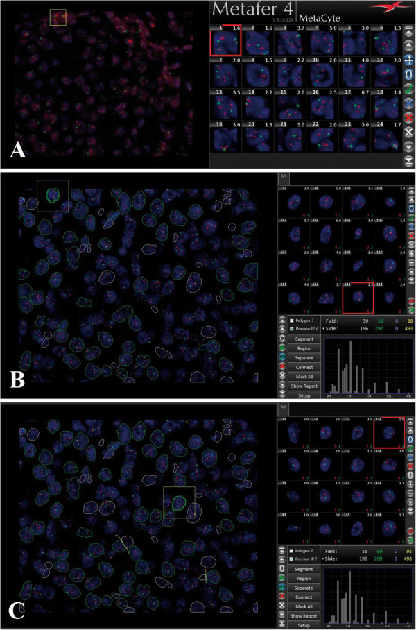
Amplification of the human epidermal growth factor receptor 2 (HER2) is a prognostic marker for poor clinical outcome and a predictive marker for therapeutic response to targeted therapies in breast cancer patients. With the introduction of anti-HER2 therapies, accurate assessment of HER2 status has become essential. Fluorescence in situ hybridization (FISH) is a widely used technique for the determination of HER2 status in breast cancer. However, the manual signal enumeration is time-consuming. Therefore, several companies like MetaSystem have developed automated image analysis software. Some of these signal enumeration software employ the so called "tile-sampling classifier", a programming algorithm through which the software quantifies fluorescent signals in images on the basis of square tiles of fixed dimensions. Considering that the size of tile does not always correspond to the size of a single tumor cell nucleus, some users argue that this analysis method might not completely reflect the biology of cells. For that reason, MetaSystems has developed a new classifier which is able to recognize nuclei within tissue sections in order to determine the HER2 amplification status on nuclei basis. We call this new programming algorithm "nuclei-sampling classifier". In this study, we evaluated the accuracy of the "nuclei-sampling classifier" in determining HER2 gene amplification by FISH in nuclei of breast cancer cells. To this aim, we randomly selected from our cohort 64 breast cancer specimens (32 nonamplified and 32 amplified) and we compared results obtained through manual scoring and through this new classifier. The new classifier automatically recognized individual nuclei. The automated analysis was followed by an optional human correction, during which the user interacted with the software in order to improve the selection of cell nuclei automatically selected. Overall concordance between manual scoring and automated nuclei-sampling analysis was 98.4% (100% for nonamplified cases and 96.9% for amplified cases). However, after human correction, concordance between the two methods was 100%. We conclude that the nuclei-based classifier is a new available tool for automated quantitative HER2 FISH signals analysis in nuclei in breast cancer specimen and it can be used for clinical purposes.
Figures

References
-
- Gianni L, Dafni U, Gelber RD, Azambuja E, Muehlbauer S, Goldhirsch A, Untch M, Smith I, Baselga J, Jackisch C. et al. Treatment with trastuzumab for 1 year after adjuvant chemotherapy in patients with HER2-positive early breast cancer: a 4-year follow-up of a randomised controlled trial. Lancet Oncol. 2011;12(3):236–244. doi: 10.1016/S1470-2045(11)70033-X. - DOI - PubMed
-
- Hanna W, O’Malley FP, Barnes P, Berendt R, Gaboury L, Magliocco A, Pettigrew N, Robertson S, Sengupta S, Tetu B. et al. Updated recommendations from the Canadian National Consensus Meeting on HER2/neu testing in breast cancer. Curr Oncol. 2007;14(4):149–153. doi: 10.3747/co.2007.131. - DOI - PMC - PubMed
Publication types
MeSH terms
Substances
LinkOut - more resources
Full Text Sources
Other Literature Sources
Medical
Research Materials
Miscellaneous
