

Sclerosing rhabdomyosarcoma presenting in the masseter muscle: a case report
- PMID: 23379991
- PMCID: PMC3573915
- DOI: 10.1186/1746-1596-8-18
Sclerosing rhabdomyosarcoma presenting in the masseter muscle: a case report
Abstract
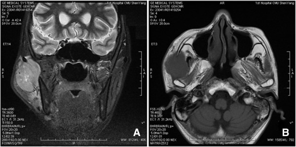
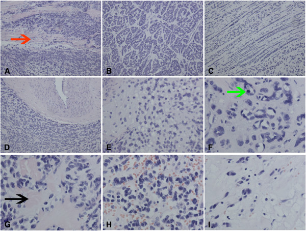
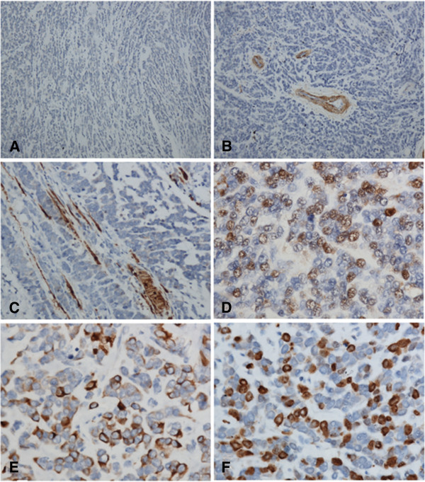
Sclerosing rhabdomyosarcoma (SRMS) is exceedingly rare, and may cause a great diagnostic confusion. Histologically, it is characterized by abundant extracellular hyalinized matrix mimicking primitive chondroid or osteoid tissue. So, it may be easily misdiagnosed as chondrosarcoma, osteosarcoma, angiosarcoma and so on. Herein, we report a case of SRMS occurring in the masseter muscle in a 40-year-old male. The tumor showed a diverse histological pattern. The tumor cells were arranged into nests, cords, pseudovascular, adenoid, microalveoli and even single-file arrays. Immunostaining showed that the tumor was positive for the Vimentin, Desmin and MyoD1, and was negative for CK, P63, NSE, CD45, CD30, S-100, CD99, Myoglobin, CD68, CD34, CD31, and α-SMA. Based on the morphological finding and immunostaining, it was diagnosed as a SRMS. In addition, focally, our case also displayed a cribriform pattern resembling adenoid cystic carcinoma. This may represent a new histological feature which can broaden the histological spectrum of this tumor and also may lead to diagnostic confusion.
Virtual slides: The virtual slide(s) for this article can be found here: http://www.diagnosticpathology.diagnomx.eu/vs/1615846455818924.
Figures
Similar articles
-
Sclerosing rhabdomyosarcoma: presentation of a rare sarcoma mimicking myoepithelial carcinoma of the parotid gland and review of the literature.Head Neck Pathol. 2015 Mar;9(1):147-52. doi: 10.1007/s12105-014-0540-x. Epub 2014 Apr 8. Head Neck Pathol. 2015. PMID: 24710732 Free PMC article. Review.
-
Sclerosing rhabdomyosarcoma in adults: report of four cases of a hyalinizing, matrix-rich variant of rhabdomyosarcoma that may be confused with osteosarcoma, chondrosarcoma, or angiosarcoma.Am J Surg Pathol. 2002 Sep;26(9):1175-83. doi: 10.1097/00000478-200209000-00008. Am J Surg Pathol. 2002. PMID: 12218574
-
[Sclerosing rhabdomyosarcoma: a clinicopathologic study of four cases with review of literature].Zhonghua Bing Li Xue Za Zhi. 2007 Sep;36(9):587-91. Zhonghua Bing Li Xue Za Zhi. 2007. PMID: 18070445 Review. Chinese.
-
The co-expression of cytokeratin and p63 in epithelioid angiosarcoma of the parotid gland: a diagnostic pitfall.Diagn Pathol. 2012 Sep 3;7:118. doi: 10.1186/1746-1596-7-118. Diagn Pathol. 2012. PMID: 22943673 Free PMC article.
-
Spindle cell/sclerosing rhabdomyosarcoma: case series from a single institution emphasizing morphology, immunohistochemistry and follow-up.Int J Clin Exp Pathol. 2015 Nov 1;8(11):13814-20. eCollection 2015. Int J Clin Exp Pathol. 2015. PMID: 26823695 Free PMC article.
Cited by
-
Spindle cell/sclerosing rhabdomyosarcoma with intracranial invasion without destroying the bone of the skull base: a case report and literature review.Acta Radiol Open. 2017 Aug 18;6(8):2058460117727316. doi: 10.1177/2058460117727316. eCollection 2017 Aug. Acta Radiol Open. 2017. PMID: 28839951 Free PMC article.
-
Head and Neck Rhabdomyosarcoma: Clinical and Pathologic Characterization of Seven Cases.Head Neck Pathol. 2017 Sep;11(3):321-326. doi: 10.1007/s12105-016-0771-0. Epub 2016 Nov 28. Head Neck Pathol. 2017. PMID: 27896667 Free PMC article.
-
Sclerosing rhabdomyosarcoma: presentation of a rare sarcoma mimicking myoepithelial carcinoma of the parotid gland and review of the literature.Head Neck Pathol. 2015 Mar;9(1):147-52. doi: 10.1007/s12105-014-0540-x. Epub 2014 Apr 8. Head Neck Pathol. 2015. PMID: 24710732 Free PMC article. Review.
-
Low-grade myxofibrosarcoma following a metal implantation in femur: a case report.Diagn Pathol. 2014 Jan 20;9:6. doi: 10.1186/1746-1596-9-6. Diagn Pathol. 2014. PMID: 24444015 Free PMC article.
-
A functional variant on 20q13.33 related to glioma risk alters enhancer activity and modulates expression of multiple genes.Hum Mutat. 2021 Jan;42(1):77-88. doi: 10.1002/humu.24134. Epub 2020 Nov 22. Hum Mutat. 2021. PMID: 33169458 Free PMC article.
References
-
- Fletcher CDM, Unni KK, Mertens F. Skeletal muscle tumors. Lyon: IARC Press; 2006. pp. 141–154. (WHO classification of tumours: pathology and genetics of tumours of soft tissue and bone. 3rd ed).
-
- Folpe AL, McKenney JK, Bridge JA. et al.Sclerosing rhabdomyosarcoma in adults: report of four cases of a hyalinizing, matrix-rich variant of rhabdomyosarcoma that may be confused with osteosarcoma, chondrosarcoma, or angiosarcoma. Am J Surg Pathol. 2002;26:1175. doi: 10.1097/00000478-200209000-00008. - DOI - PubMed
-
- Vadgama B, Sebire NJ, Malone M. et al.Sclerosing rhabdomyosarcoma in childhood: case report and review of the literature. Pediatr Dev Pathol. 2004;7:391. - PubMed
Publication types
MeSH terms
Substances
LinkOut - more resources
Full Text Sources
Other Literature Sources
Medical
Research Materials
Miscellaneous
