. 2013 Apr;57(4):1823-31.
doi: 10.1128/AAC.02237-12. Epub 2013 Feb 4.

Rattusin, an intestinal α-defensin-related peptide in rats with a unique cysteine spacing pattern and salt-insensitive antibacterial activities

Affiliations

Rattusin, an intestinal α-defensin-related peptide in rats with a unique cysteine spacing pattern and salt-insensitive antibacterial activities

Amar A Patil et al. Antimicrob Agents Chemother. 2013 Apr.

Abstract

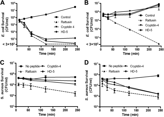
Cationic antimicrobial peptides are essential components of the innate immune system. As a major family of mammalian antimicrobial peptides, defensins are expressed mainly by mucosal epithelial cells and promyelocytes. Despite the capacity to kill a broad spectrum of bacteria through physical disruption of membranes, most defensins show substantially reduced antibacterial activities in the presence of monovalent and divalent cations, thereby limiting their therapeutic potential, particularly for the treatment of systemic infections. Genome-wide computational screening of the rat genome led to the identification of the gene for a novel α-defensin-related peptide that we termed rattusin. Rattusin shares a highly conserved signal and prosequence with mammalian α-defensins, but instead of the canonical α-defensin six-cysteine motif, rattusin consists of five cysteines with a distinctive spacing pattern. Furthermore, rattusin is preferentially expressed in Paneth cells of the distal small intestine with potent antibacterial activity against a broad range of Gram-negative and Gram-positive bacteria, including antibiotic-resistant strains. The MICs were mostly in the range of 2 to 4 μM, with no appreciable toxicity to mammalian cells at up to 100 μM. In contrast to classical α- and β-defensins, rattusin retained its activity in the presence of physiological concentrations of NaCl and Mg(2+), making it an attractive antimicrobial candidate for both topical and systemic applications.


Figures

Fig 1
Fig 1
Schematic drawing of the structure of a mammalian defensin precursor. Although all classical α-, β-, and θ-defensins contain six cysteines with different disulfide arrays, three subfamilies of α-defensin-related sequences (defa-rs) with a different number and spacing pattern of cysteines have been found in intestinal Paneth cells of mice (CRS1C and CRS4C) and rats (rattusin). The prosequences of defensins are highly conserved within but not between subfamilies, except for β-defensins, whose prosequences are variable as well. Biologically active, mature sequences of defensins are released from proforms through proteolytic cleavage.
Fig 2
Fig 2
Amino acid sequence alignment of rattusin with representative α-defensins and related peptides in rodents and humans. Dashes were inserted to maximize the alignment. Conserved amino acids are shaded, and mature sequences are underlined. Vertical arrows indicate the known start sites of mature peptides. The length of each mature peptide is also indicated. The signal peptides and prosequences of the peptides are conserved, whereas carboxyl-terminal mature peptides are diversified. Note that the spacing pattern of cysteine residues in rattusin differs from those of all other defensins and defensin-related peptides. Abbreviations: rDefa6, rat α-defensin 6; RatNP4, rat neutrophil peptide α-defensin 4; DEFA5/HD5, human α-defensin 5.
Fig 3
Fig 3
mRNA expression patterns of rattusin, MMP7, and three isoforms of trypsin in the rat gastrointestinal tract by RT-PCR. The housekeeping gene for GAPDH was used to normalize template input. Prox., proximal; Mid., middle.
Fig 4
Fig 4
Localization of rattusin mRNA in rat ileal crypts. In situ hybridization was performed with rat ileal sections probed with rattusin-specific, DIG-labeled antisense (A) and sense (B) RNA probes, followed by anti-DIG antibody reaction and color development. Note the positive staining throughout the base of intestinal crypts (indicated by arrows) in panel A but not in panel B.
Fig 5
Fig 5
RP-HPLC profile of reduced and oxidized rattusin. The reduced synthetic peptide was refolded by air oxidation. Refolded rattusin was purified to homogeneity by RP-HPLC. Note that there is an approximately 5.5-min decrease in the retention time of oxidized rattusin due to refolding.
Fig 6
Fig 6
Antibacterial activities of reduced and oxidized forms of rattusin in the presence or absence of 100 mM NaCl. A colony counting assay was conducted following 2 h of incubation of bacteria with each peptide in serial 2-fold dilutions in duplicate. (A) Activity against E. coli ATCC 25922 in the absence of NaCl. (B) Activity against E. coli ATCC 25922 in the presence of NaCl. (C) Activity against S. aureus ATCC 25923 in the absence of NaCl. (D) Activity against S. aureus ATCC 25923 in the presence of NaCl. Data shown are the means ± the standard errors of the means of a representative of two independent experiments.
Fig 7
Fig 7
Kinetics of killing of E. coli and S. aureus in the presence or absence of 100 mM NaCl by rattusin, Crp4, and HD5. E. coli O157:H7 ATCC 700728 was incubated with 4 μM rattusin, Crp4, or HD5 or an equal volume of 0.01% acetic acid (control) in duplicate with (A) or without 100 mM NaCl (B) for 10, 30, 60, 120, or 240 min. S. aureus ATCC 25923 was incubated with or without each peptide at 2 μM in duplicate in the absence (C) or presence (D) of 100 mM NaCl. Surviving bacteria were plated and counted. Data shown are the means ± the standard errors of the means of two independent experiments.
Fig 8
Fig 8
Effect of salinity on the antibacterial activity of rattusin. E. coli O157:H7 ATCC 700728 (A, C) and S. aureus ATCC 25923 (B, D) were incubated with 4 and 2 μM rattusin, respectively, with increasing concentrations of NaCl (A, B) or MgCl2 (C, D) for 4 h. Surviving bacteria were plated and counted. Data shown are the means ± the standard errors of the means of two or three independent experiments.
Fig 9
Fig 9
Absence of cytotoxicity of rattusin and Crp4 to Caco-2 cells. Cells were incubated with 100 μM rattusin, Crp4, or fowlicidin-1 in duplicate for 24 h with or without 10% FBS. Cell viability was measured by an alamarBlue dye-based method. The data shown are representative of two independent experiments.

References

    1. Finch RG. 2004. Antibiotic resistance: a view from the prescriber. Nat. Rev. Microbiol. 2:989–994 - PubMed
    1. Zasloff M. 2002. Antimicrobial peptides of multicellular organisms. Nature 415:389–395 - PubMed
    1. Hancock RE, Sahl HG. 2006. Antimicrobial and host-defense peptides as new anti-infective therapeutic strategies. Nat. Biotechnol. 24:1551–1557 - PubMed
    1. Brogden KA. 2005. Antimicrobial peptides: pore formers or metabolic inhibitors in bacteria? Nat. Rev. Microbiol. 3:238–250 - PubMed
    1. Selsted ME, Ouellette AJ. 2005. Mammalian defensins in the antimicrobial immune response. Nat. Immunol. 6:551–557 - PubMed

Publication types

MeSH terms