

MMP-2 suppression abrogates irradiation-induced microtubule formation in endothelial cells by inhibiting αvβ3-mediated SDF-1/CXCR4 signaling
- PMID: 23381805
- PMCID: PMC3586295
- DOI: 10.3892/ijo.2013.1806
MMP-2 suppression abrogates irradiation-induced microtubule formation in endothelial cells by inhibiting αvβ3-mediated SDF-1/CXCR4 signaling
Abstract
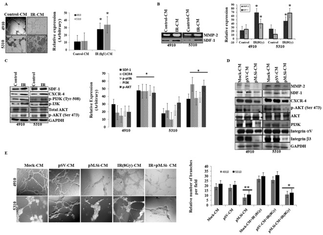
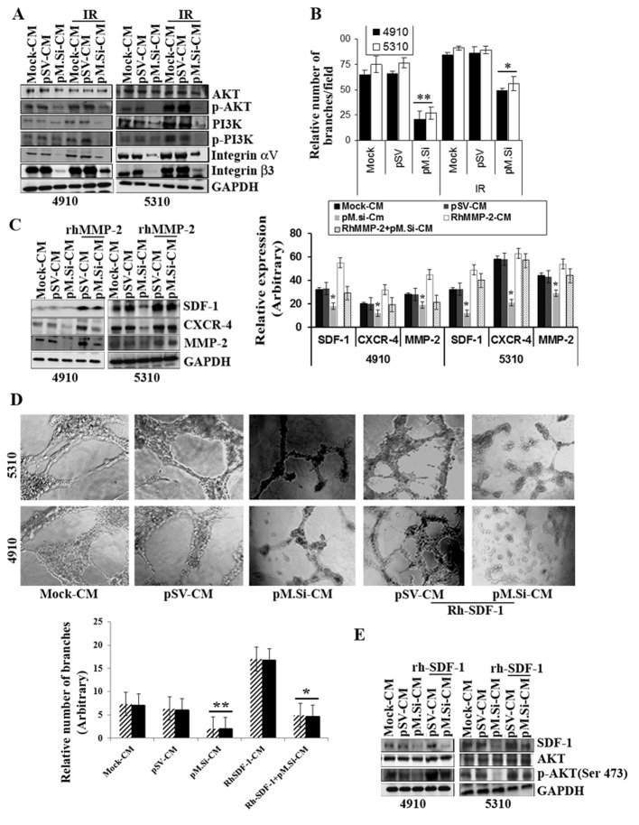
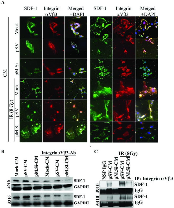
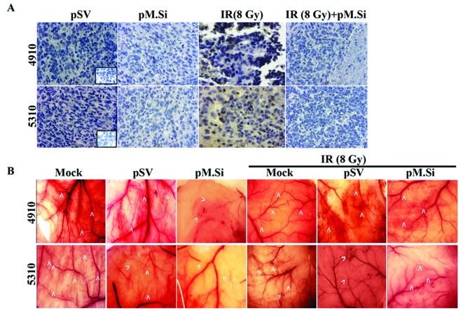
The majority of glioblastoma multiforme (GBM) tumors recur after radiation (IR) treatment due to increased angiogenesis and IR-induced signaling events in endothelial cells (ECs) that are involved in tumor neovascularization; however, these signaling events have yet to be well characterized. In the present study, we observed that IR (8 Gy) significantly elevated MMP-2 expression and gelatinolytic activity in 4910 and 5310 human GBM xenograft cells. In addition, ECs treated with tumor-conditioned media (CM) obtained from IR-treated 4910 and 5310 cells showed increased microtubule formation. In view of this finding, we investigated the possible anti-angiogenic effects of MMP-2 downregulation using siRNA (pM.si) in IR-treated cells. We also determined the effect of CM obtained from mock, pSV (scrambled vector) and pMMP-2.si on endothelial cell growth and vessel formation. pM.si-CM-treated ECs showed inhibited IR-CM-induced SDF-1, CXCR4, phospho-PI3K and phospho-AKT and αvβ3 expression levels. In vitro angiogenesis assays also showed that the pM.si+IR decreased IR-induced vessel formation in ECs. Immunofluorescence and immunoprecipitation experiments indicated the abrogation of αvβ3-SDF-1 interaction in pM.si-CM-treated ECs when compared to mock or pSV treatments. External supplementation of either rhMMP-2 or rhSDF-1 counteracted and noticeably reversed pM.si-inhibited SDF-1, CXCR4, phospho-PI3K and phospho-AKT expression levels and angiogenesis, thereby confirming the role of MMP-2 in the regulation of αvβ3-mediated SDF-1/CXCR4 signaling. In addition to the in vitro results, the in vivo mouse dorsal air sac model also showed reduced angiogenesis after injection of pM.si alone or in combination with IR-treated xenograft cells. In contrast, injection of mock or pSV-treated cells resulted in robust formation of characteristic neovascularization. Collectively, our data demonstrate the role of MMP-2 in the regulation of SDF-1/CXCR4 signaling-mediated angiogenesis in ECs and show the anti-angiogenic efficacy of combining MMP-2 downregulation and IR when treating patients with GBM in the future.
Figures
Similar articles
-
Tissue inhibitor of metalloproteinase 3 suppresses tumor angiogenesis in matrix metalloproteinase 2-down-regulated lung cancer.Cancer Res. 2008 Jun 15;68(12):4736-45. doi: 10.1158/0008-5472.CAN-07-6612. Cancer Res. 2008. PMID: 18559520 Free PMC article.
-
Osteopontin enhances the expression and activity of MMP-2 via the SDF-1/CXCR4 axis in hepatocellular carcinoma cell lines.PLoS One. 2011;6(8):e23831. doi: 10.1371/journal.pone.0023831. Epub 2011 Aug 31. PLoS One. 2011. PMID: 21909361 Free PMC article.
-
MMP-2 alters VEGF expression via alphaVbeta3 integrin-mediated PI3K/AKT signaling in A549 lung cancer cells.Int J Cancer. 2010 Sep 1;127(5):1081-95. doi: 10.1002/ijc.25134. Int J Cancer. 2010. PMID: 20027628 Free PMC article.
-
Targeting SDF-1/CXCR4 to inhibit tumour vasculature for treatment of glioblastomas.Br J Cancer. 2011 Jun 7;104(12):1805-9. doi: 10.1038/bjc.2011.169. Epub 2011 May 17. Br J Cancer. 2011. PMID: 21587260 Free PMC article. Review.
-
Vasculogenesis: a crucial player in the resistance of solid tumours to radiotherapy.Br J Radiol. 2014 Mar;87(1035):20130686. doi: 10.1259/bjr.20130686. Br J Radiol. 2014. PMID: 24338942 Free PMC article. Review.
Cited by
-
Effect of integrin α5β1 inhibition on SDF-l/CXCR4-mediated choroidal neovascularization.Int J Ophthalmol. 2018 May 18;11(5):726-735. doi: 10.18240/ijo.2018.05.04. eCollection 2018. Int J Ophthalmol. 2018. PMID: 29862169 Free PMC article.
-
Effects of RNA silencing of matrix metalloproteinase-2 on the growth of esophageal carcinoma cells in vivo.Oncol Lett. 2017 Mar;13(3):1119-1124. doi: 10.3892/ol.2016.5542. Epub 2016 Dec 27. Oncol Lett. 2017. PMID: 28454222 Free PMC article.
-
Cisplatin targets the stromal cell-derived factor-1-CXC chemokine receptor type 4 axis to suppress metastasis and invasion of ovarian cancer-initiating cells.Tumour Biol. 2014 May;35(5):4637-44. doi: 10.1007/s13277-014-1607-8. Epub 2014 Jan 10. Tumour Biol. 2014. PMID: 24408020
-
Critical role of aberrant angiogenesis in the development of tumor hypoxia and associated radioresistance.Cancers (Basel). 2014 Apr 8;6(2):813-28. doi: 10.3390/cancers6020813. Cancers (Basel). 2014. PMID: 24717239 Free PMC article.
-
Inhibition of MMP-2 Expression Enhances the Antitumor Effect of Sorafenib in Hepatocellular Carcinoma by Suppressing the PI3K/AKT/mTOR Pathway.Oncol Res. 2017 Nov 2;25(9):1543-1553. doi: 10.3727/096504017X14886444100783. Epub 2017 Mar 8. Oncol Res. 2017. PMID: 28276313 Free PMC article.
References
-
- Ribatti D, Djonov V. Angiogenesis in development and cancer today. Int J Dev Biol. 2011;55:343–344. - PubMed
-
- Kargiotis O, Geka A, Rao JS, Kyritsis AP. Effects of irradiation on tumor cell survival, invasion and angiogenesis. J Neurooncol. 2010;100:323–338. - PubMed
-
- Wachsberger P, Burd R, Dicker AP. Tumor response to ionizing radiation combined with antiangiogenesis or vascular targeting agents: exploring mechanisms of interaction. Clin Cancer Res. 2003;9:1957–1971. - PubMed
Publication types
MeSH terms
Substances
Grants and funding
LinkOut - more resources
Full Text Sources
Other Literature Sources
Molecular Biology Databases
Miscellaneous
