. 2013 Apr;33(8):1503-14.
doi: 10.1128/MCB.01716-12. Epub 2013 Feb 4.

IDOL stimulates clathrin-independent endocytosis and multivesicular body-mediated lysosomal degradation of the low-density lipoprotein receptor

Affiliations

IDOL stimulates clathrin-independent endocytosis and multivesicular body-mediated lysosomal degradation of the low-density lipoprotein receptor

Elena Scotti et al. Mol Cell Biol. 2013 Apr.

Abstract

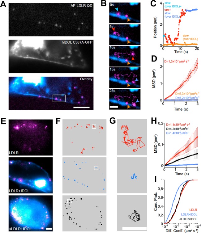
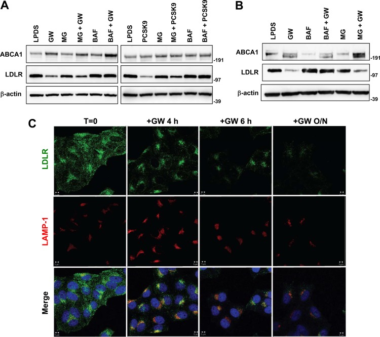
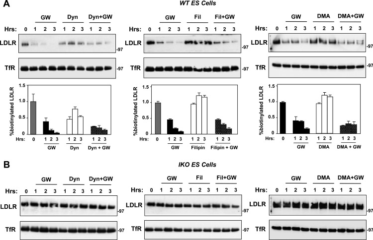
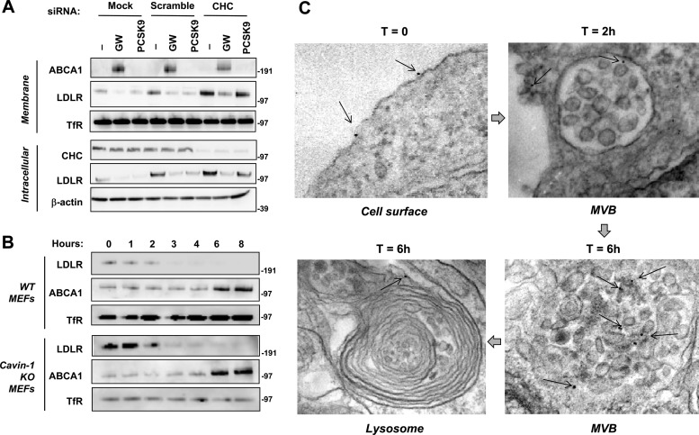
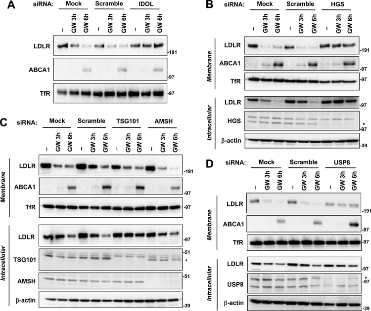
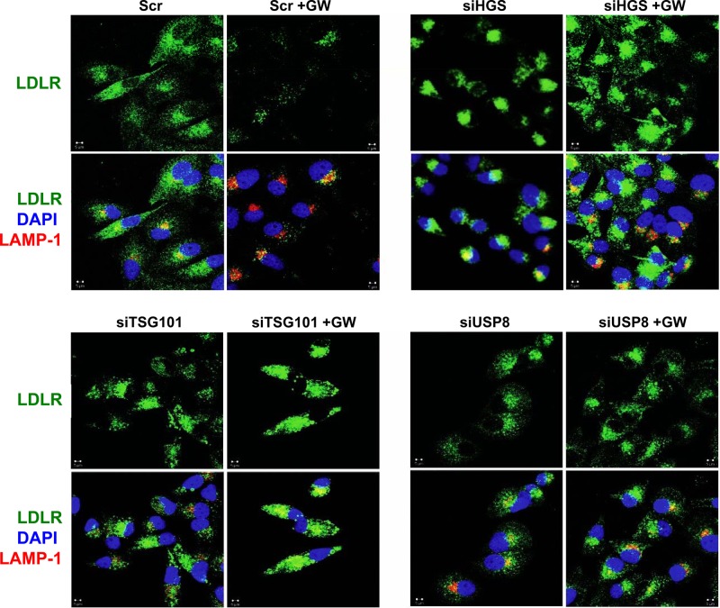
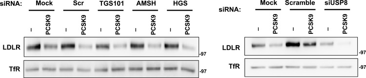
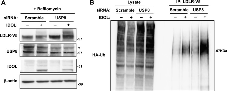
The low-density lipoprotein receptor (LDLR) is a critical determinant of plasma cholesterol levels that internalizes lipoprotein cargo via clathrin-mediated endocytosis. Here, we show that the E3 ubiquitin ligase IDOL stimulates a previously unrecognized, clathrin-independent pathway for LDLR internalization. Real-time single-particle tracking and electron microscopy reveal that IDOL is recruited to the plasma membrane by LDLR, promotes LDLR internalization in the absence of clathrin or caveolae, and facilitates LDLR degradation by shuttling it into the multivesicular body (MVB) protein-sorting pathway. The IDOL-dependent degradation pathway is distinct from that mediated by PCSK9 as only IDOL employs ESCRT (endosomal-sorting complex required for transport) complexes to recognize and traffic LDLR to lysosomes. Small interfering RNA (siRNA)-mediated knockdown of ESCRT-0 (HGS) or ESCRT-I (TSG101) components prevents IDOL-mediated LDLR degradation. We further show that USP8 acts downstream of IDOL to deubiquitinate LDLR and that USP8 is required for LDLR entry into the MVB pathway. These results provide key mechanistic insights into an evolutionarily conserved pathway for the control of lipoprotein receptor expression and cellular lipid uptake.


Figures

Fig 1
Fig 1
The LXR-IDOL pathway blocks LDL association with LDLR at the plasma membrane in living cells. (A) HepG2 cells stably overexpressing GFP-LDLR were treated with GW3965 (1 μM) overnight and then incubated with DiI-LDL. The association of DiI-LDL was determined in living cells by spinning-disc confocal microscopy. Still images at time zero and 2.5 h are shown. The entire movie shown in Video S1 in the supplemental material covers 3 h. (B) Immunoblot analysis of IDOL protein expression in transiently transfected 293T cells with monoclonal antibody (10E7F10). Cells were treated with or without MG132 for 4 h prior to harvest. (C) Immunoblot analysis of endogenous IDOL protein expression with antibody 10E7F10 in wild-type (WT) and IDOL-deficient cells. Primary mouse peritoneal macrophages were starved in lipoprotein-deficient medium and then treated with GW3965 overnight and MG132 for 5 h. T, time.
Fig 2
Fig 2
LDLR recruits IDOL to the plasma membrane in a FERM-dependent manner. T-Rex cells were transfected with BirA-ER and the indicated constructs and then labeled with streptavidin-Alexa Fluor 568 (10 μg/ml), fixed, and stained with V5 antibody. Nuclei were counterstained with DAPI (blue). Representative confocal images are shown. Fluorescence profiles from the sections indicated by the arrows are shown in the lower panels. (A) Localization of AP-LDLR with IDOL C387A-GFP. (B) Localization of AP-LDLR with IDOL C387A-V5. (C) Localization of AP-K6R K20R C29A LDLR (K6/K20R C29A LDLR) with IDOL-V5. (D) Localization of AP-LDLR with ΔFERM IDOL-GFP. (E) Localization of AP-LDLR with FERM-GFP. (F) Localization of AP-Δ792 LDLR with IDOL-GFP.
Fig 3
Fig 3
IDOL induces clustering and restricts the mobility of LDLR in the plasma membrane. Single-particle tracking using QDs was used to study the interaction between LDLR and IDOL. (A) Living T-Rex cells cotransfected for 36 h with AP-LDLR, BirA-ER, and IDOL-GFP, pretreated with tetracycline and biotin, and surface-labeled with streptavidin-QDs emitting at 655 nm. Scale bar, 5 μm. (B) Magnification of the white region in panel A shows real-time changes in the trajectory of an LDLR molecule in relation to IDOL submembrane clusters. Scale bar, 1 μm. (C to D) Position vector and MSD analysis of the LDLR molecule indicated by colored arrows in panel B. Colors correspond to changes in LDLR mobility, which are dependent on the interaction with IDOL. The MSD was calculated over the time indicated according to the colors in panel C. (E) Examples of cells overexpressing AP-LDLR alone, AP-LDLR and IDOL-GFP, or AP-Δ792 LDLR and IDOL-GFP. Scale bar, 2 μm. (F and G) QD-labeled LDLR trajectories corresponding to images in panel E. Scale bar, 1 μm. (H) The cumulative probability distributions of diffusion coefficients showing decreased mobility of LDLR in cells cotransfected with IDOL-GFP. No significant differences were observed between cells expressing LDLR alone and AP-Δ792 LDLR together with IDOL.
Fig 4
Fig 4
IDOL induces late-endosome/lysosome localization of LDLR coincident with degradation. (A) HepG2 cells were treated with GW3965 (GW; 1 μM) or PCSK9 (5 μg/ml) for 1 h, and then proteasome inhibitor (25 μM MG132 [MG]) or lysosome inhibitor (50 nM bafilomycin [Baf]) was added for an additional 5 h. The levels of ABCA1 and LDLR were determined by immunoblotting. Similar results were obtained in three independent experiments. Numbers to the right of the blots are molecular weight markers. (B) HeLa cells were processed and analyzed as described for panel A. (C) HeLa cells were cultured in 10% LPDS medium for 8 h and then treated with GW3965 (1 μM) for the indicated times. Cells were immunostained with LDLR and LAMP-1 antibodies. Nuclei were counterstained with DAPI (blue). Representative confocal images are shown. T, time; O/N, overnight.
Fig 5
Fig 5
IDOL induces LDLR internalization independent of clathrin, caveolae, or macropinocytosis. (A) Wild-type (WT) ES cells were grown in sterol depletion medium (10% LPDS with 5 μM simvastatin and 100 μM mevalonic acid) for 16 h. Cells were then pretreated with GW3965 (1 μM) for 1 h, and subsequently dynasore (Dyn; 80 μM), filipin (Fil; 1 μg/ml), or 5-(N,N-dimethyl) amiloride hydrochloride (DMA; 100 μM) was added for the indicated times. Surface proteins were collected and analyzed as described in Materials and Methods. Results from replicate experiments are quantitated below each blot. (B) IDOL-null (IKO) ES cells were analyzed as described for panel A.
Fig 6
Fig 6
IDOL stimulates LDLR internalization through a clathrin- and caveola-independent pathway. (A) HeLa cells were transfected with siRNA targeting CHC (clathrin heavy chain) or a universal control siRNA or the transfection reagent only (mock) for 96 h as described in Materials and Methods. Then the cells were cultured in 10% LPDS medium for 8 h and treated with GW3965 (1 μM) or PCSK9 (5 μg/ml) overnight. Expression of ABCA1, LDLR, and CHC was assessed by immunoblotting. Endogenous transferrin receptor (TfR) and actin levels were also determined as loading controls for membrane and intracellular lysates, respectively. (B) Wild-type and cavin-1 KO MEFs were cultured in 10% LPDS medium for 16 h and then treated with GW3965 (1 μM) for the indicated times. Membrane lysates were analyzed by immunoblotting. Similar results were obtained in two independent experiments. (C) Visualization of IDOL-dependent LDLR trafficking to MVBs and lysosomes. HepG2 cells were transduced with an AP-Y807C LDLR expression vector along with control or tetracycline-inducible IDOL expression vectors. After 12 h of serum, AP-Y807C LDLR was labeled with biotin. Cells were then treated with doxycycline to induce IDOL and labeled with streptavidin gold beads for 2 to 6 h. Representative electron micrographs are shown.
Fig 7
Fig 7
IDOL facilitates LDLR degradation by shuttling it into the MVB protein-sorting pathway. (A to D) HeLa cells were transfected with indicated siRNAs for 48 or 96 h as indicated in Materials and Methods, followed by GW3965 (1 μM) treatment in lipoprotein-deficient medium for the indicated times. Membrane and intracellular cell lysates were prepared and analyzed by immunoblotting. Efficiency of knockdown was confirmed by immunoblotting using polyclonal anti-HGS (B), anti-TSG101 (C), anti-AMSH (C), and anti-USP8 (D) antibodies. Endogenous transferrin receptor (TfR) and actin levels were also measured as controls for membrane and intracellular lysates, respectively. Blots are representative of three independent experiments. *, specific band.
Fig 8
Fig 8
Silencing of the MVB pathway blocks IDOL-mediated LDLR degradation and localization of the LDLR in late endosomes/lysosomes. HeLa cells were transfected with the indicated siRNAs for 48 or 96 h, followed by GW3965 (GW; 1 μM) treatment in lipoprotein-deficient medium for 6 h. Cells were then fixed and stained with LDLR and LAMP-1 antibodies. Nuclei were counterstained with DAPI (blue). Representative confocal images are shown. Scr, scrambled siRNA; siHGS, siRNA targeting HGS; siTSG101, siRNA targeting TSG101; siUSP8, siRNA targeting USP8.
Fig 9
Fig 9
Inactivation of MVB pathway does not affect PCSK9-driven LDLR degradation. HeLa cells were transfected with the indicated siRNA for 48 or 96 h as described in the legend of Fig. 7, followed by PCSK9 (5 μg/ml) or BSA (Ctrl) treatment in lipoprotein-deficient medium for 12 h. Membrane cell lysates were prepared, and proteins were analyzed by immunoblotting.
Fig 10
Fig 10
USP8 is required for deubiquitination of LDLR downstream of IDOL action in the MVB pathway. (A) 293T cells were transfected with a control siRNA (scramble) or siRNA targeting USP8. After 24 h, the cells were transfected with V5-tagged LDLR, HA-tagged ubiquitin (HA-Ub), and human IDOL expression vectors. The siRNA transfection was then repeated after 24 h, and bafilomycin was added to the medium for 6 h. Total lysates were analyzed by immunoblotting. *, specific band. (B) V5-tagged LDLR in the cell lysates was immunoprecipitated (IP) using anti-V5 antibody followed by immunoblotting for HA-ubiquitin. Blots are representative of two independent experiments.

Similar articles

Cited by

References

    1. Russell DW, Schneider WJ, Yamamoto T, Luskey KL, Brown MS, Goldstein JL. 1984. Domain map of the LDL receptor: sequence homology with the epidermal growth factor precursor. Cell 37:577–585 - PubMed
    1. Brown MS, Goldstein JL. 1986. A receptor-mediated pathway for cholesterol homeostasis. Science 232:34–47 - PubMed
    1. Tolleshaug H, Hobgood KK, Brown MS, Goldstein JL. 1983. The LDL receptor locus in familial hypercholesterolemia: multiple mutations disrupt transport and processing of a membrane receptor. Cell 32:941–951 - PubMed
    1. Hua X, Yokoyama C, Wu J, Briggs MR, Brown MS, Goldstein JL, Wang X. 1993. SREBP-2, a second basic-helix-loop-helix-leucine zipper protein that stimulates transcription by binding to a sterol regulatory element. Proc. Natl. Acad. Sci. U. S. A. 90:11603–11607 - PMC - PubMed
    1. Zelcer N, Hong C, Boyadjian R, Tontonoz P. 2009. LXR regulates cholesterol uptake through Idol-dependent ubiquitination of the LDL receptor. Science 325:100–104 - PMC - PubMed

Publication types

MeSH terms

Substances

LinkOut - more resources