. 2013 Feb 19;110(8):E716-25.
doi: 10.1073/pnas.1215707110. Epub 2013 Feb 4.

Ikaros promotes early-born neuronal fates in the cerebral cortex

Affiliations

Ikaros promotes early-born neuronal fates in the cerebral cortex

Jessica M Alsiö et al. Proc Natl Acad Sci U S A. .

Erratum in

Abstract

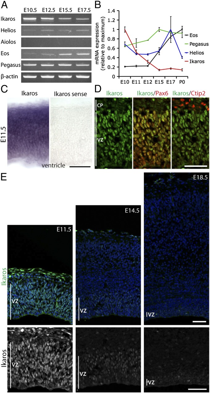
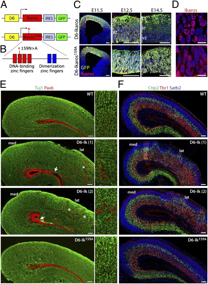
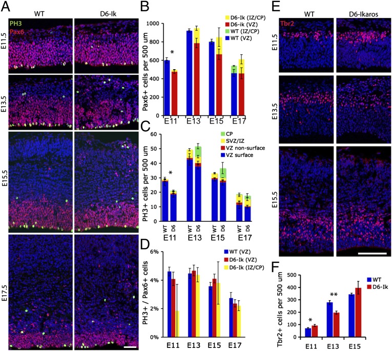
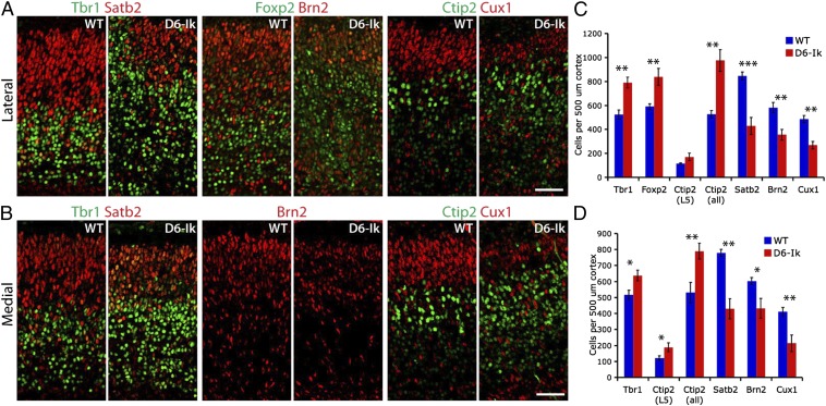
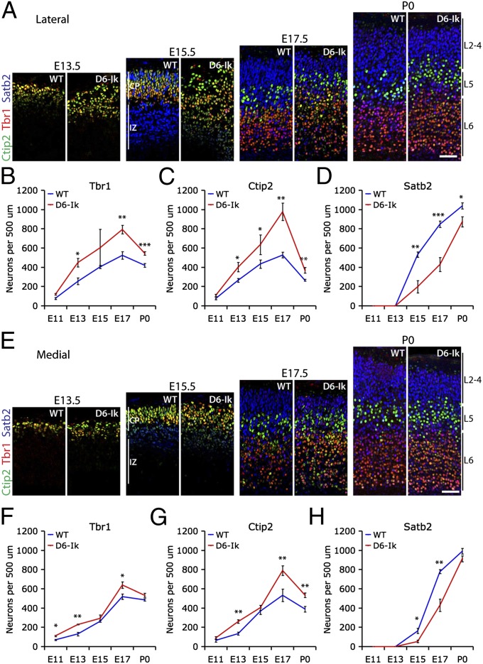
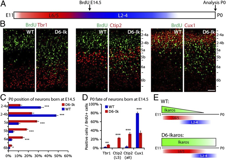
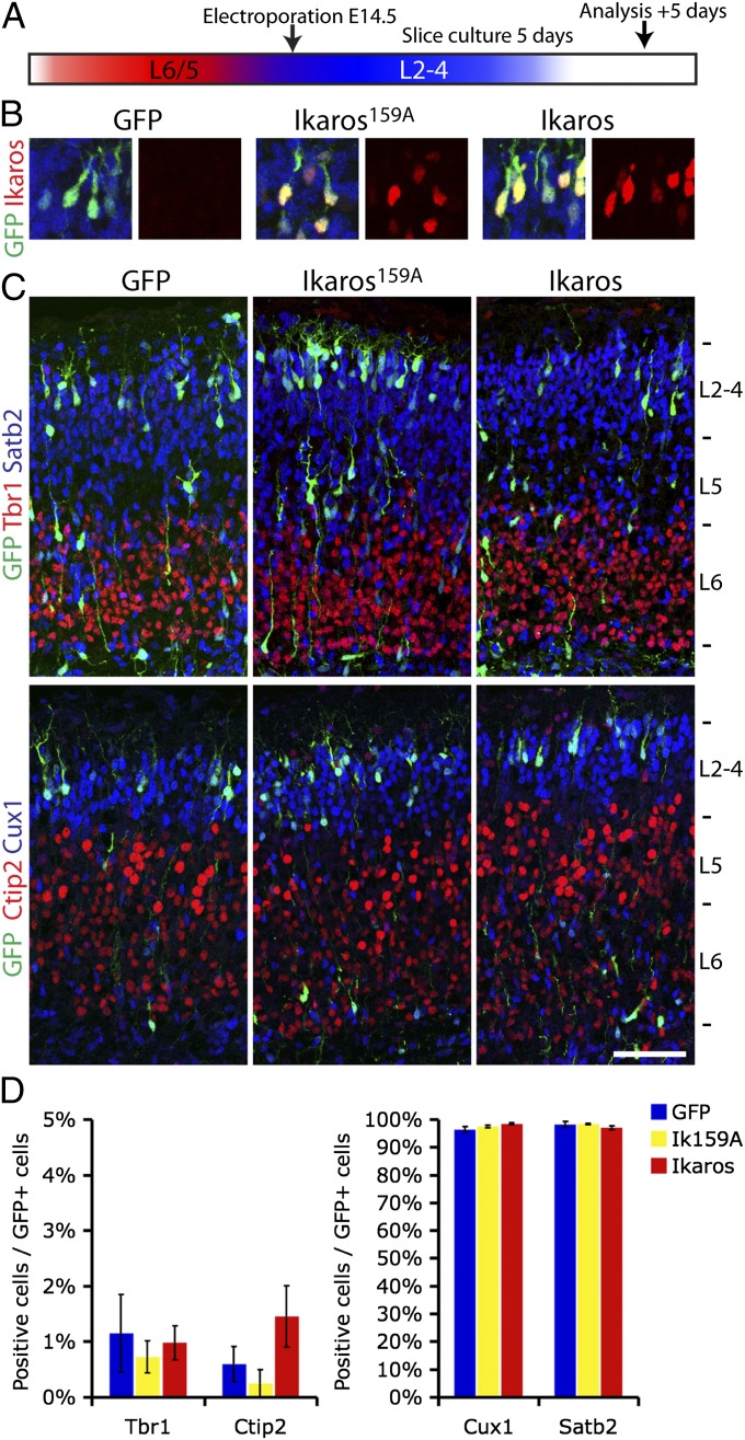
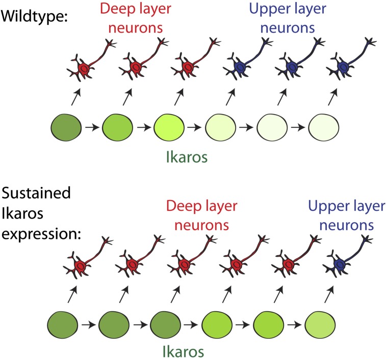
During cerebral cortex development, a series of projection neuron types is generated in a fixed temporal order. In Drosophila neuroblasts, the transcription factor hunchback encodes first-born identity within neural lineages. One of its mammalian homologs, Ikaros, was recently reported to play an equivalent role in retinal progenitor cells, raising the question as to whether Ikaros/Hunchback proteins could be general factors regulating the development of early-born fates throughout the nervous system. Ikaros is also expressed in progenitor cells of the mouse cerebral cortex, and this expression is highest during the early stages of neurogenesis and thereafter decreases over time. Transgenic mice with sustained Ikaros expression in cortical progenitor cells and neurons have developmental defects, including displaced progenitor cells within the cortical plate, increased early neural differentiation, and disrupted cortical lamination. Sustained Ikaros expression results in a prolonged period of generation of deep layer neurons into the stages when, normally, only late-born upper layer neurons are generated, as well as a delayed production of late-born neurons. Consequently, more early-born and fewer late-born neurons are present in the cortex of these mice at birth. This phenotype was observed in all parts of the cortex, including those with minimal structural defects, demonstrating that it is not secondary to abnormalities in cortical morphogenesis. These data suggest that Ikaros plays a similar role in regulating early temporal fates in the mammalian cerebral cortex as Ikaros/Hunchback proteins do in the Drosophila nerve cord.


Conflict of interest statement

The authors declare no conflict of interest.

Figures

Fig. 1.
Fig. 1.
Ikaros is expressed in cortical progenitor cells at high levels early, decreasing over developmental time. (A) Semiquantitative RT-PCR for Ikaros family members in cortex of different developmental stages. Ikaros, Helios, Eos, and Pegasus are expressed in the developing cortex, but Aiolos is not. (B) Real-time qRT-PCR for Ikaros, Helios, Eos, and Pegasus in cortex from E10.5 to P0 shows that Ikaros mRNA levels decrease over developmental time in the cortex. Expression was normalized to the average expression of six housekeeping genes (GAPDH, β-actin, TBP, UBC, YWHAZ, and SDHA). For each gene, values are shown relative to the time point with the highest expression level and represent the mean normalized expression ± SEM (n = 3 independent RNA extractions). (C) In situ hybridization for Ikaros in E11.5 mouse cortex shows specific signal throughout the cortical wall that was not detected with a sense probe. (D and E) Immunostaining for Ikaros (green) in mouse cortical sections at different stages of development, as indicated. (D) At the beginning of neurogenesis, at E11.5, Ikaros is expressed in virtually all Pax6+ cortical progenitor cells of the VZ but not in neurons of the cortical plate (CP), as identified by Ctip2 staining. (E) At mid- (E14.5) and late (E18.5) neurogenic stages, Ikaros is still detected in cortical progenitor cells in the VZ but at much lower levels than at E11.5. All sections in E were processed in parallel from dissection through to imaging. (Scale bars: 50 μm.)
Fig. 2.
Fig. 2.
Ikaros-expressing progenitor cells generate projection neurons of all laminar fates. (A and B) BAC-Ikaros-Cre transgenic mouse crossed to the R26-YFP reporter mouse was used for lineage analysis of neurons produced from Ikaros-expressing progenitor cells. A low recombination rate gives rise to distinguishable clones of progenitor cells and their progeny at E13.5. (CF) In the adult brain, progeny of Ikaros-Cre–expressing progenitor cells (YFP+) were mainly neurons (NeuN+) and found throughout the layers of the cortex. (D and E) YFP+ progeny in deep cortical layers frequently expressed Foxp2 and Tbr1, two transcription factors expressed in layer 6 projection neurons. (F) YFP+ neurons in the upper layers were often positive for Cux1, a marker of cortical projection neurons in layers 2–3. (Scale bars: AF, 100 μm; D′–F′, 50 μm.)
Fig. 3.
Fig. 3.
Increased Ikaros expression in the cortex affects cortical architecture and is dependent on Ikaros DNA binding ability. (A and B) Constructs used for generation of D6-Ikaros and D6-Ikaros159A transgenic mice. (A) Ikaros and Ikaros159A followed by IRES-GFP were cloned downstream of the D6 enhancer sequence, which drives expression specifically in the cortex and hippocampus. (B) Ikaros159A protein has an asparagine-to-alanine point mutation in the second DNA-binding zinc finger, which disrupts its ability to bind DNA (41). (C) Sections of D6-Ikaros (Upper) and D6-Ikaros159A (Lower) cortex at different developmental stages stained for GFP and Ikaros. Both transgenes are expressed specifically in the cortex. GFP and Ikaros expression from the D6-Ikaros transgene, but not from the D6-Ikaros159A transgene, is down-regulated in the VZ and IZ starting at E12.5. (D) In the D6-Ikaros cortex, Ikaros protein is localized to pericentromeric foci (Upper), whereas in D6-Ikaros159A, the Ikaros159A protein is more diffusely localized throughout the nucleus (Lower). (E) Sections of WT, two lines of D6-Ikaros, and one line of D6-Ikaros159A P0 cortex immunostained for neurons (Tuj1, green) and progenitor cells (Pax6, red). In WT and D6-Ikaros159A cortex, the large majority of Pax6+ progenitor cells are confined to the VZ. In D6-Ikaros brains, ectopic Pax6+ cells were also found scattered throughout the cortical wall (asterisks and Insets). In severely affected D6-Ikaros brains, the integrity of the VZ was also disrupted (arrows). This phenotype was more severe in the lateral cortex (lat), whereas the most medial cortex (med) and hippocampus displayed a grossly normal structure. (F) Sections of WT, two lines of D6-Ikaros, and one line of D6-Ikaros159A P0 cortex stained for transcription factors expressed in neurons of specific cortical layers: Tbr1 (layer 6 neurons, red), Ctip2 (layer 5 neurons, green), and Satb2 (layer 2–4 neurons, blue). D6-Ikaros, but not D6-Ikaros159A, displays a disorganized laminar structure and an increased width of layer 6. (Scale bars: C and EF, 100 μm; D, 10 μm.)
Fig. 4.
Fig. 4.
Ectopic progenitor cells and proliferation in the Ikaros-overexpressing cortex. (A) Immunostaining for Pax6+ radial glial progenitor cells and the mitotic marker pH3 in WT and D6-Ikaros cortex shows that ectopic Pax6 progenitor cells are present in the cortical plate (CP) and IZ throughout developmental stages E11.5–E17.5 in the D6-Ikaros cortex. (B) Total number of Pax6+ progenitor cells (including both VZ-bound and ectopic progenitors) was significantly lower in D6-Ikaros at E11.5 but normal from E13.5–E17.5 (n = 3–4 brains). (C) More mitotic (ph3+) cells are found in the cortical plate/IZ of D6-Ikaros compared with WT littermates throughout the stages. The total number of mitotic cells is decreased in D6-Ikaros at E11.5 but unaffected at later stages (n = 3–4 brains). SVZ, subventricular zone. (D) Mitotic index of Pax6+ progenitor cells is not affected in D6-Ikaros cortex (n = 3–4 brains). (E and F) Numbers of Tbr2+ IPCs in D6-Ikaros compared with WT cortex were increased at E11.5, decreased at E13.5, and indistinguishable at E15.5 (n = 3 brains; *P < 0.05; **P < 0.01). (Scale bars: A, 50 μm; E, 100 μm.)
Fig. 5.
Fig. 5.
More deep layer neurons and fewer upper layer neurons following sustained Ikaros expression in the cortex. (A and B) Immunostaining for deep layer-specific transcription factors Tbr1 (layer 6), Foxp2 (layer 6), and Ctip2 (weak in layer 6, strong in layer 5), and late-born, upper layer-specific transcription factors Satb2 (layers 2–4), Brn2 (layers 2–5), and Cux1 (layers 2–3) in E17.5 WT and D6-Ikaros lateral (A) and medial (B) cortex. (C and D) Significantly more early-born, deep layer neurons and significantly fewer late-born, upper layer neurons were observed in D6-Ikaros than in WT littermates, both in the lateral (C) and medial (D) parts of the cortex. Values represent mean ± SEM (n = 4 brains). *P < 0.05; **P < 0.01; ***P < 0.001. (Scale bars: 50 μm.)
Fig. 6.
Fig. 6.
Early and sustained increase in deep layer genesis and delayed upper layer genesis following sustained Ikaros expression in the cortex. (AD) Immunostaining and quantifications of Tbr1 (layer 6, red), Ctip2 (weak in layer 6 and strong in layer 5, green), and Satb2 (layers 2–4; blue) in WT and D6-Ikaros lateral cortex at different stages of cortical development as indicated. (EH) Immunostaining and quantifications of the same markers in WT and D6-Ikaros medial cortex. Increased numbers of Tbr1+ and Ctip2+ deep layer neurons were observed in D6-Ikaros compared with WT littermates throughout development both laterally and medially, as well as a delayed appearance and decreased numbers of Satb2+ upper layer neurons throughout development. In BD and FH, values represent mean ± SEM (n = 3–7 brains). *P < 0.05; **P < 0.01; ***P < 0.001. (Scale bars: 50 μm.)
Fig. 7.
Fig. 7.
BrdU birth-dating shows an extended period of deep layer neuron production following sustained Ikaros expression in the cortex. (A) Birth-dating experimental design. BrdU was administered to pregnant females at E14.5 and incorporated by cycling progenitor cells. Laminar positions and neuronal fates of strongly BrdU+ neurons born at E14.5 were analyzed at P0. (B and C) Cells born at E14.5 in the WT cortex mainly populate the upper layers (Cux1+), and very few are found in the deeper layers (Tbr1+ and Ctip2+). Cells born at E14.5 in the D6-Ikaros cortex are found at deeper positions, within the Tbr1+ and Ctip2+ layers, and also in Cux1+ upper layers. (Scale bar: B, 50 μm.) (D) In the WT cortex, fewer than 5% of neurons born at E14.5 express deep layer markers Tbr1 or Ctip2, and most (80%) express upper layer marker Cux1. In D6-Ikaros, the proportion of deep layer neurons produced at E14.5 was greatly increased (10-fold for Ctip2) and the proportion of upper layer neurons was less than half of the WT level. In C and D, values represent mean ± SEM (n = 4 brains). *P < 0.05; **P < 0.01; ***P < 0.001. (E) Schematic drawing summarizes the D6-Ikaros developmental timing phenotype. In WT cortex, Ikaros levels decrease in progenitor cells over time. Sustained Ikaros expression beyond its normal period leads to prolonged and increased production of deep layer neurons coupled to delayed production of upper layer neurons.
Fig. 8.
Fig. 8.
Reintroduction of Ikaros in late progenitor cells is not sufficient to induce heterochronically early fates. (A) Electroporation experimental design. Ikaros-ires-GFP, inactive Ikaros159A-IRES-GFP, and GFP only were electroporated into VZ progenitors of the lateral ventricle at E14.5. Brains were cultured as slices for 5 d before fates of GFP+ neurons produced from these progenitor cells were analyzed by immunohistochemistry. (B) Ikaros-IRES-GFP and Ikaros159A-IRES-GFP constructs, but not the IRES-GFP control construct, express Ikaros in GFP-positive neurons after 5 d of culture. (C and D) No differences in fates of neurons born from Ikaros and control electroporated progenitor cells were observed. In all conditions, less than 2% of neurons were expressing deep layer markers Tbr1 and Ctip2 and most neurons (>95%) were upper layer neurons expressing Cux1 and Satb2. In C, values represent mean ± SEM (n = 3 brains; 100–300 cells per brain counted). (Scale bar: C, 100 μm.)
Fig. P1.
Fig. P1.
Cortical progenitor cells (circles) generate deep-layer neurons (red) early in development, followed by upper-layer neurons (blue) late in development. (Upper) We show that progenitor cells express Ikaros (green) at early time points when they are producing deep-layer neurons. (Lower) Sustained expression of Ikaros in cortical progenitor cells beyond its normal period results in a prolonged and increased production of deep-layer neurons coupled to a delayed onset of and decreased production of upper-layer neurons.

Similar articles

Cited by

References

    1. Livesey FJ, Cepko CL. Vertebrate neural cell-fate determination: Lessons from the retina. Nat Rev Neurosci. 2001;2(2):109–118. - PubMed
    1. McConnell SK, Kaznowski CE. Cell cycle dependence of laminar determination in developing neocortex. Science. 1991;254(5029):282–285. - PubMed
    1. Frantz GD, McConnell SK. Restriction of late cerebral cortical progenitors to an upper-layer fate. Neuron. 1996;17(1):55–61. - PubMed
    1. Arlotta P, et al. Neuronal subtype-specific genes that control corticospinal motor neuron development in vivo. Neuron. 2005;45(2):207–221. - PubMed
    1. Alcamo EA, et al. Satb2 regulates callosal projection neuron identity in the developing cerebral cortex. Neuron. 2008;57(3):364–377. - PubMed

Publication types

Substances

LinkOut - more resources