. 2013 Feb 19;110(8):2940-5.
doi: 10.1073/pnas.1222798110. Epub 2013 Feb 4.

Essential requirement for IRF8 and SLC15A4 implicates plasmacytoid dendritic cells in the pathogenesis of lupus

Affiliations

Essential requirement for IRF8 and SLC15A4 implicates plasmacytoid dendritic cells in the pathogenesis of lupus

Roberto Baccala et al. Proc Natl Acad Sci U S A. .

Abstract

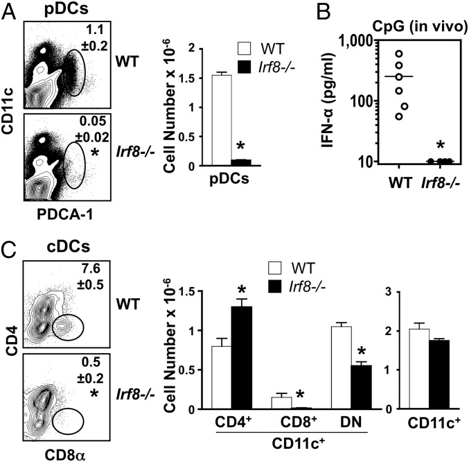
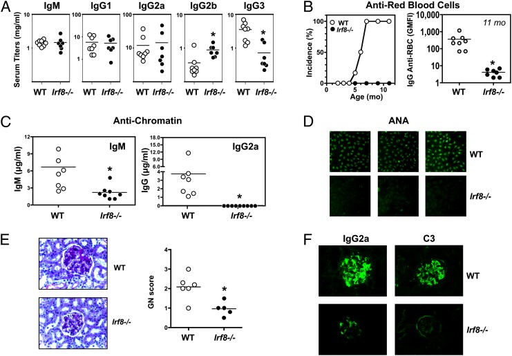
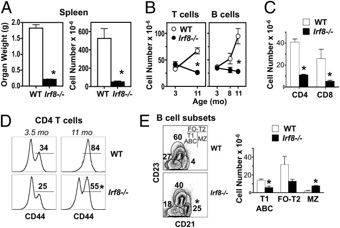
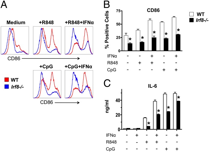
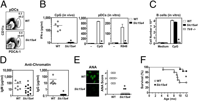
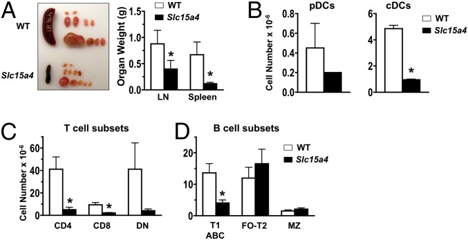
In vitro evidence suggests that plasmacytoid dendritic cells (pDCs) are intimately involved in the pathogenesis of lupus. However, it remains to be determined whether these cells are required in vivo for disease development, and whether their contribution is restricted to hyperproduction of type I IFNs. To address these issues, we created lupus-predisposed mice lacking the IFN regulatory factor 8 (IRF8) or carrying a mutation that impairs the peptide/histidine transporter solute carrier family 15, member 4 (SLC15A4). IRF8-deficient NZB mice, lacking pDCs, showed almost complete absence of anti-nuclear, anti-chromatin, and anti-erythrocyte autoantibodies, along with reduced kidney disease. These effects were observed despite normal B-cell responses to Toll-like receptor (TLR) 7 and TLR9 stimuli and intact humoral responses to conventional T-dependent and -independent antigens. Moreover, Slc15a4 mutant C57BL/6-Fas(lpr) mice, in which pDCs are present but unable to produce type I IFNs in response to endosomal TLR ligands, also showed an absence of autoantibodies, reduced lymphadenopathy and splenomegaly, and extended survival. Taken together, our results demonstrate that pDCs and the production of type I IFNs by these cells are critical contributors to the pathogenesis of lupus-like autoimmunity in these models. Thus, IRF8 and SLC15A4 may provide important targets for therapeutic intervention in human lupus.


Conflict of interest statement

The authors declare no conflict of interest.

Figures

Fig. 1.
Fig. 1.
Absence of pDCs and CD8α+ cDCs in young IRF8-deficient NZB mice. Mutant and WT NZB mice (age 3 mo; n = 3–6/group) were analyzed for cellular differences in spleen and in vivo responses to CpG challenge. (A) pDC frequency and numbers. Spleen cells were assessed by flow cytometry using anti-CD11c and anti-PDCA-1 antibodies. Similar results were obtained when cells were stained with antibodies to B220, SiglecH, and CD11b (data not shown). (B) In vivo type I IFN production in response to TLR9 engagement. Serum IFN-α levels in CpG-challenged mice were determined by ELISA. (C) cDC frequency and numbers. Spleen cells were analyzed by flow cytometry after gating on CD11c+ cells to identify CD4+, CD8+, and CD4CD8 cDC subsets. Numbers within the flow cytometry plots correspond to average frequencies of the indicated subsets. Error bars in graphs indicate SD. Asterisks indicate statistical significance (P < 0.05).
Fig. 2.
Fig. 2.
Reduced autoimmunity in IRF8-deficient NZB mice. Mutant and WT NZB mice (age 11 mo; n = 5–8/group) were examined for serologic and histological disease characteristics. (A) Polyclonal serum Ig levels. (B) Anti-erythrocyte autoantibodies. Individuals with a geometric mean fluorescence intensity >50 were considered positive for anti-RBC autoantibodies. (C) Anti-chromatin autoantibodies. (D) ANAs, with representative individuals shown. (E) Representative PAS-stained kidney sections and glomerulonephritis scores. (F) Kidney deposits (IgG2a and C3), with representative immunofluorescence images shown. Asterisks indicate statistical significance (P < 0.05).
Fig. 3.
Fig. 3.
Reduced T-cell and B-cell expansion in older IRF8-deficient NZB mice. Mutant and WT NZB mice (age 8–11 mo; n = 3/group) were analyzed for cellular changes in spleen and peritoneal cavity. (A) Spleen weight and cellularity at 11 mo. (B) T-cell and B-cell numbers in spleen at age 11 mo. Cells were analyzed by flow cytometry after gating on TCRβ+ (T cell) or B220+ (B cell) populations. (C) CD4+ and CD8+ T-cell subsets in spleen at age 11 mo. (D) Frequency of activated CD4+ T cells in spleen at age 11 mo. Gated CD4+ T cells were assessed for expression of CD44. (E) B-cell subsets in spleen at age 8 mo. Gated B220+IgM+ cells were analyzed for transitional T2 and follicular (T2-FO), marginal zone (MZ), and T1 immature and age-associated (T1-ABC) B cells. Numbers within flow cytometry plots correspond to average frequencies of the indicated subsets. Error bars in graphs indicate SD. Asterisks indicate statistical significance (P < 0.05).
Fig. 4.
Fig. 4.
Defective in vitro responses by cDCs from IRF8-deficient NZB mice. cDCs were differentiated from BM cells from WT and mutant mice (age 3 mo; pools of 2–3 mice) in the presence of GM-CSF, then stimulated with endosomal TLR ligands in the presence or absence of IFN-α. (A and B) CD86 up-regulation defined by flow cytometry after gating on CD11c+ cells. (C) IL-6 production determined by ELISA. One representative of two independent experiments is shown. Error bars in graphs indicate SD of samples analyzed in triplicate. Asterisks indicate statistical significance (P < 0.05).
Fig. 5.
Fig. 5.
Reduced autoimmunity in Slc15a4 mutant C57BL/6-Faslpr mice. Mutant and WT mice were analyzed for pDC development and function at age 4 mo (n = 3–5/group), disease manifestations at age 8 mo (n = 7–10/group), and survival (n = 10–13/group). (A) Frequency of pDCs in spleen. Cells were assessed by flow cytometry using anti-CD11c and anti–PDCA-1 antibodies. Similar results were obtained using antibodies to B220, SiglecH, and CD11b (data not shown). (B) In vivo and in vitro type I IFN production in response to endosomal TLR stimulation. Serum IFN-α levels in CpG-challenged mice, and IFN-α production by BM-differentiated pDCs stimulated with CpG or R848, were determined by ELISA. (C) In vitro B-cell proliferation in response to TLR9 engagement. Purified B cells from mutant and WT mice were stimulated in vitro with CpG. B cells from Tlr9−/− C57BL/6 mice were used as negative controls. (D) Anti-chromatin autoantibodies. (E) ANA. (F) Survival. Error bars in graphs indicate SD. Asterisks indicate statistical significance (P < 0.05).
Fig. 6.
Fig. 6.
Cellular changes in Slc15a4 mutant C57BL/6-Faslpr mice. Spleen and LNs of mutant and WT mice were examined at age 8 mo (n = 3/group). (A) Spleen and LN organ weights. (B) pDC and cDC numbers in spleen. Flow cytometry was used to identify pDCs (PDCA-1+CD11clow) and cDCs (CD11c+). (C) T-cell numbers in spleen. CD4+, CD8+, and CD4CD8 (double-negative) T cells were identified by flow cytometry after gating on TCRβ+ cells. (D) B-cell numbers in spleen. Spleen cells were examined by flow cytometry as in Fig 3E. Error bars in graphs indicate SD. Asterisks indicate statistical significance (P < 0.05).

References

    1. Baechler EC, Gregersen PK, Behrens TW. The emerging role of interferon in human systemic lupus erythematosus. Curr Opin Immunol. 2004;16(6):801–807. - PubMed
    1. Kirou KA, et al. Activation of the interferon-alpha pathway identifies a subgroup of systemic lupus erythematosus patients with distinct serologic features and active disease. Arthritis Rheum. 2005;52(5):1491–1503. - PubMed
    1. Obermoser G, Pascual V. The interferon-alpha signature of systemic lupus erythematosus. Lupus. 2010;19(9):1012–1019. - PMC - PubMed
    1. Santiago-Raber ML, et al. Type-I interferon receptor deficiency reduces lupus-like disease in NZB mice. J Exp Med. 2003;197(6):777–788. - PMC - PubMed
    1. Braun D, Geraldes P, Demengeot J. Type I interferon controls the onset and severity of autoimmune manifestations in lpr mice. J Autoimmun. 2003;20(1):15–25. - PubMed

Publication types