. 2013 Feb 19;110(8):E653-61.
doi: 10.1073/pnas.1221050110. Epub 2013 Feb 4.

Intrinsic evolutionary constraints on protease structure, enzyme acylation, and the identity of the catalytic triad

Affiliations

Intrinsic evolutionary constraints on protease structure, enzyme acylation, and the identity of the catalytic triad

Andrew R Buller et al. Proc Natl Acad Sci U S A. .

Abstract

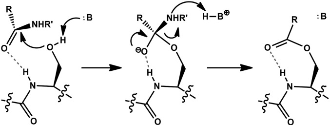
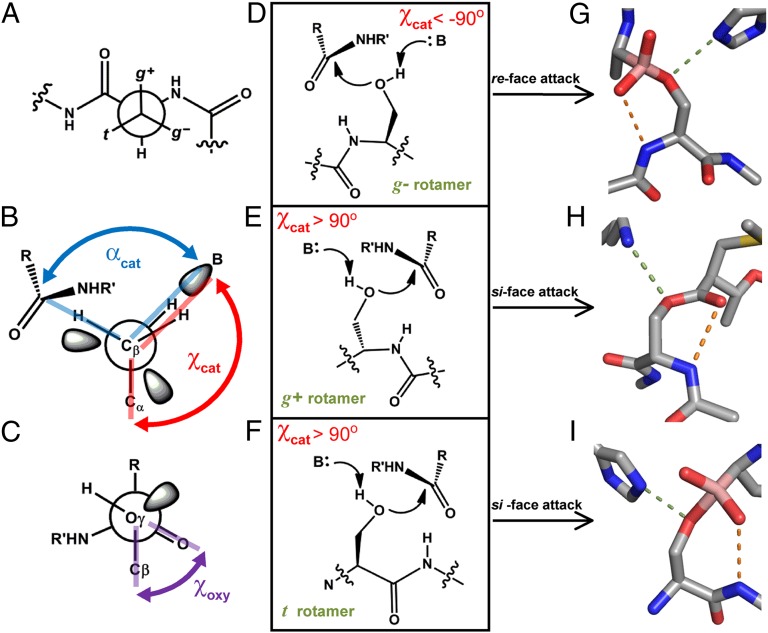
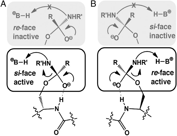
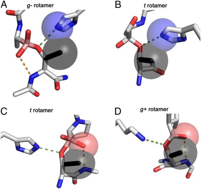
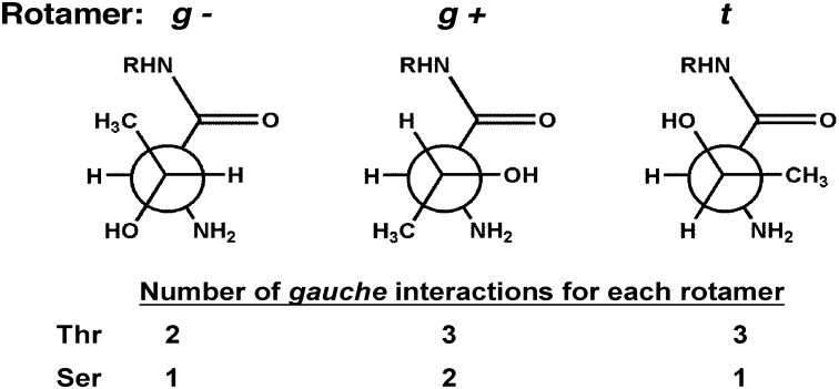
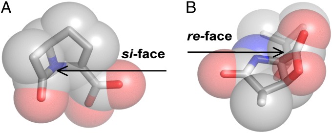
The study of proteolysis lies at the heart of our understanding of biocatalysis, enzyme evolution, and drug development. To understand the degree of natural variation in protease active sites, we systematically evaluated simple active site features from all serine, cysteine and threonine proteases of independent lineage. This convergent evolutionary analysis revealed several interrelated and previously unrecognized relationships. The reactive rotamer of the nucleophile determines which neighboring amide can be used in the local oxyanion hole. Each rotamer-oxyanion hole combination limits the location of the moiety facilitating proton transfer and, combined together, fixes the stereochemistry of catalysis. All proteases that use an acyl-enzyme mechanism naturally divide into two classes according to which face of the peptide substrate is attacked during catalysis. We show that each class is subject to unique structural constraints that have governed the convergent evolution of enzyme structure. Using this framework, we show that the γ-methyl of Thr causes an intrinsic steric clash that precludes its use as the nucleophile in the traditional catalytic triad. This constraint is released upon autoproteolysis and we propose a molecular basis for the increased enzymatic efficiency introduced by the γ-methyl of Thr. Finally, we identify several classes of natural products whose mode of action is sensitive to the division according to the face of attack identified here. This analysis of protease structure and function unifies 50 y of biocatalysis research, providing a framework for the continued study of enzyme evolution and the development of inhibitors with increased selectivity.


Conflict of interest statement

The authors declare no conflict of interest.

Figures

Fig. 1.
Fig. 1.
The acylation mechanism of a Ser protease. After substrate binding, the Ser side chain attacks the scissile peptide bond to generate a tetrahedral oxyanion intermediate. Protonation of the amine leaving group allows collapse of the intermediate and formation of an acyl-enzyme species. Dashed bonds represent the hydrogen bond of the oxyanion hole. See text for details.
Fig. 2.
Fig. 2.
Active site architectures of Ser Proteases. (A) Newman projection down the Cβ-Cα bond of a side chain on a peptide. The rotamers of the γ-atom are named g+, g−, or t, as shown. (B) Newman projection down the Cβ-Oγ bond of a Ser nucleophile. The dihedral angle χcat is measured between Cα, Cβ, Oγ, and the base (B). αcat is defined as the angle between the base, Oγ, and carbonyl carbon. (C) The dihedral angle between Cβ, Oγ of the nucleophile, and the carbonyl of the scissile bond, χoxy, is used to measure the relative orientation of the substrate. (D-F) Schematic of the three possible active site arrangements when a local oxyanion hole is used. (G–I) Representative examples of a Ser protease with each reactive rotamer. Dashes represent hydrogen bonds between the nucleophile and the base (green) or the protein backbone and substrate (orange). PDB IDs: G, 3VGC, γ-chymotrypsin with a boronic acid inhibitor; H, 2EEP, signal peptidase I with a β-lactam inhibitor; I, 1B12, prolyl aminopeptidase with a boronic acid inhibitor.
Fig. 3.
Fig. 3.
Stereospecificity of proteolysis. (A) The substrate carbonyl points toward the local backbone for stabilization with a local oxyanion hole. When χcat > 90°, the substrate must expose the si face of the peptide for attack, because a re-face attack is nonproductive for proton transfer. (B) When χcat < 90°, the opposite holds. Only a re-face attack is viable for enzyme acylation and a si-face attack leads to an inactive complex.
Fig. 4.
Fig. 4.
Steric clashes of the Thr γ-methyl. (A) γ-Chymotrypsin with a boronic acid inhibitor (PDB ID: 3VGC). The γ-methyl of Thr was modeled (black) and its steric clash with the base is indicated by overlapping spheres drawn with the van der Waals radii. (B) CMV protease with an α-ketoamide inhibitor (PDB ID: 1NKM) shows a steric clash still occurs when the local oxyanion hole is not used. (C) Prolyl aminopeptidase with a boronic acid inhibitor (PDB ID: 2EEP). The “nucleophilic elbow” motif is incompatible with the Thr γ-methyl, which clashes with the oxyanion substrate (red sphere). (D) A similar clash is observed for signal peptidase I with a β-lactam inhibitor (PDB ID: 1B12).
Fig. 5.
Fig. 5.
Gauche interactions of an N-terminal nucleophile (Ntn). The g− rotamer of an Ntn Thr has the fewest gauche interactions. When the γ-methyl is removed by mutation to Ser, the t rotamer becomes favored. This effect is exacerbated by repulsive interactions with the neighboring carbonyl. Consequently, the Thr γ-methyl shifts the rotamer distribution toward a reactive state, which increases kcat. These effects are intrinsic to Ntn structure and may contribute to the evolutionary selection of a Thr nucleophile.
Fig. 6.
Fig. 6.
Plot of the φ, ψ angles of the nucleophilic residue for Ser and Cys overlaying the Ramachandran plot. Proteases cluster according to the reactive rotamer in catalysis. The outlier, marked X, does not use the local backbone for oxyanion stabilization. Ramachandran plot was generated in MolProbity: light blue, strongly favored; dark blue, allowed (85).
Fig. 7.
Fig. 7.
Structural overlay of proteases according to their reactive rotamer. (A) Enzymes that use the g− rotamer show no structural similarity apart from their φ, ψ angles. (B) A reactive g+ rotamer has a loop at its N-terminal direction and a helix at it C-terminal end. (C) Use of the t rotamer and the N + 1 oxyanion hole imparts stringent structural constraints that are satisfied by a β-strand turning into an α-helix, the “nucleophilic elbow” motif. Structures shown are listed in Tables 1 and 2.
Fig. 8.
Fig. 8.
Stereospecific antibiotics. (A) The β-lactam molecule carbapenem contains stereochemistry that blocks the si face of the elecrophilic carbonyl and this molecule may be used only to inhibit re-face attacking enzymes. (B) Conversely, omuralide features a β-lactone–γ-lactam core that occludes the re face of the eletrophilic carbonyl. Consequently, this inhibitor scaffold is effective only against si-face–attacking enzymes.

References

    1. Warshel A, Naray-Szabo G, Sussman F, Hwang JK. How do serine proteases really work? Biochemistry. 1989;28(9):3629–3637. - PubMed
    1. Fersht A. Structure and Mechanism in Protein Science: A Guide to Enzyme Catalysis and Protein Folding. New York: Freeman; 1999.
    1. Kraut J. How do enzymes work? Science. 1988;242(4878):533–540. - PubMed
    1. Warshel A. Electrostatic origin of the catalytic power of enzymes and the role of preorganized active sites. J Biol Chem. 1998;273(42):27035–27038. - PubMed
    1. Warshel A. Energetics of enzyme catalysis. Proc Natl Acad Sci USA. 1978;75(11):5250–5254. - PMC - PubMed

Publication types