. 2013 Feb 4;200(3):287-300.
doi: 10.1083/jcb.201209113.

RabGEFs are a major determinant for specific Rab membrane targeting

Affiliations

RabGEFs are a major determinant for specific Rab membrane targeting

Julia Blümer et al. J Cell Biol. .

Abstract

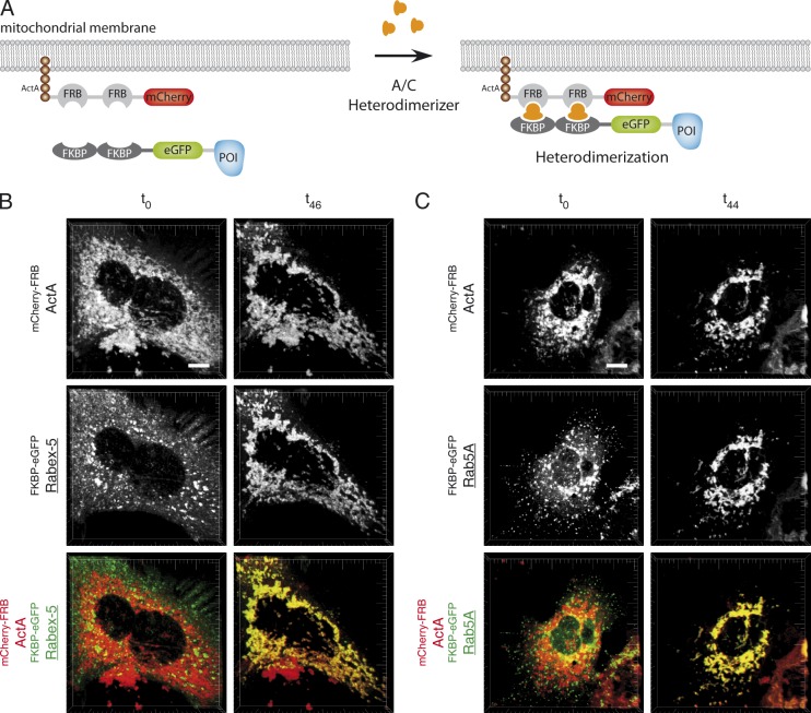
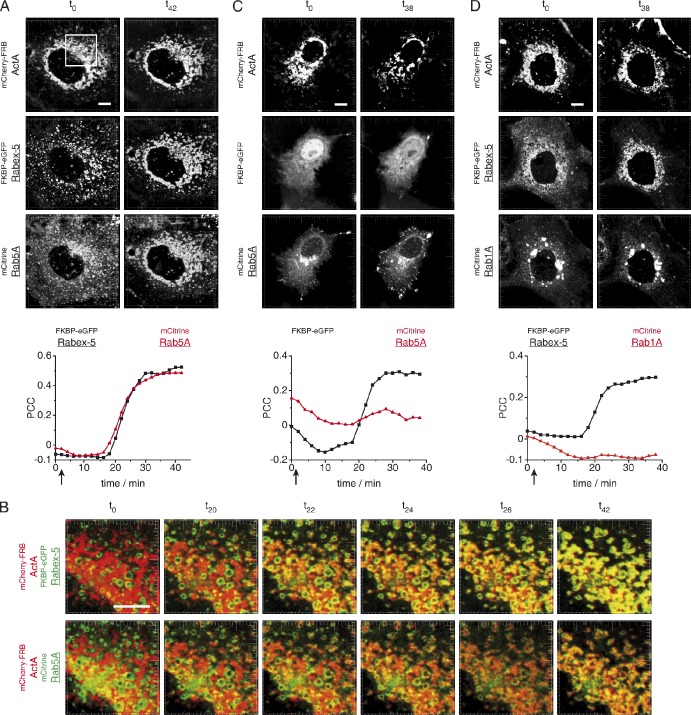
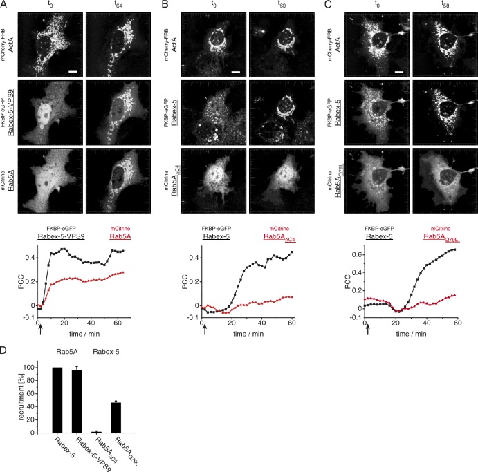
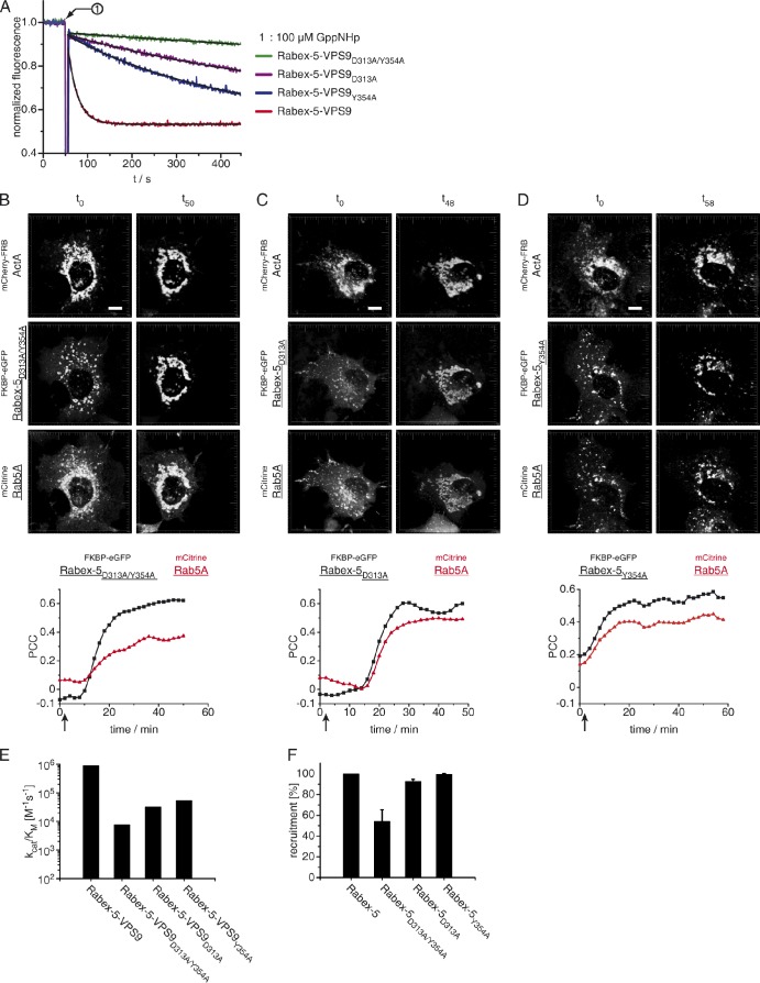
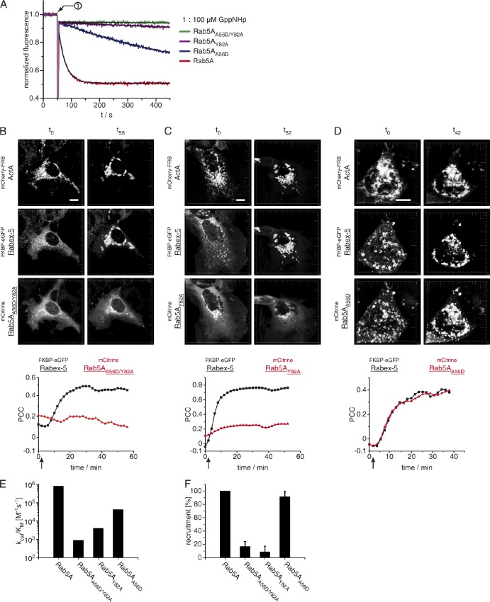
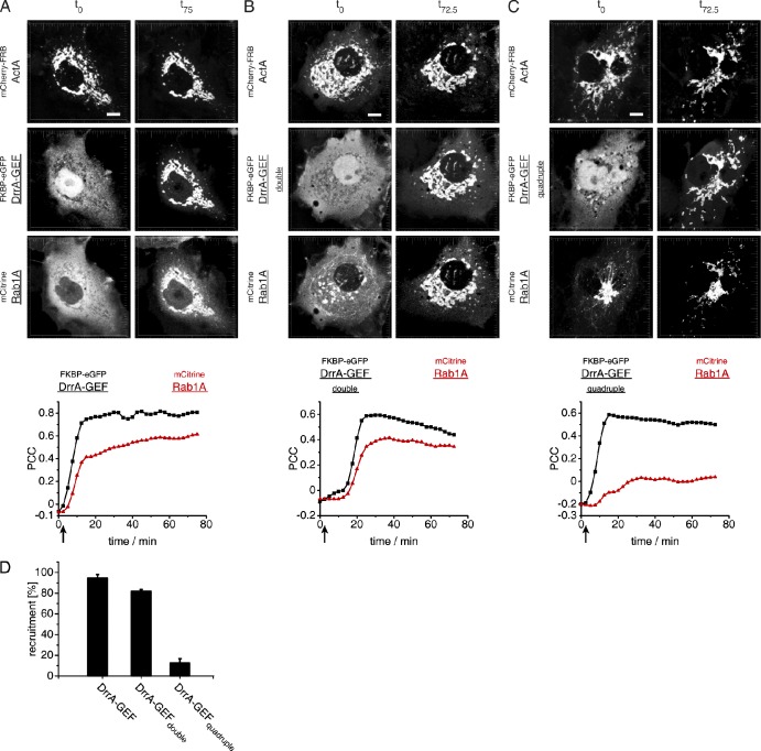
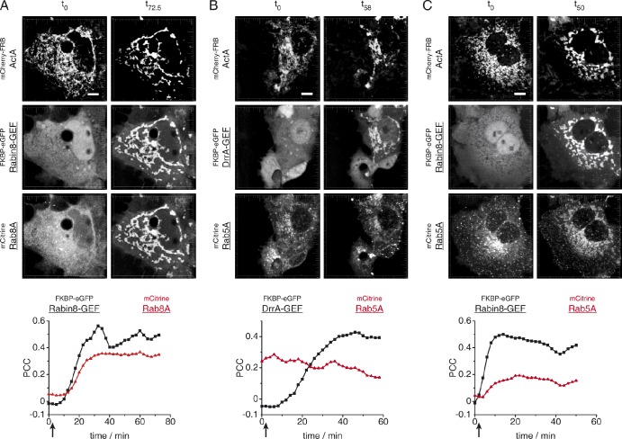
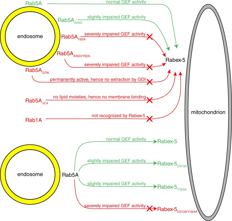
Eukaryotic cells critically depend on the correct regulation of intracellular vesicular trafficking to transport biological material. The Rab subfamily of small guanosine triphosphatases controls these processes by acting as a molecular on/off switch. To fulfill their function, active Rab proteins need to localize to intracellular membranes via posttranslationally attached geranylgeranyl lipids. Each member of the manifold Rab family localizes specifically to a distinct membrane, but it is unclear how this specific membrane recruitment is achieved. Here, we demonstrate that Rab-activating guanosine diphosphate/guanosine triphosphate exchange factors (GEFs) display the minimal targeting machinery for recruiting Rabs from the cytosol to the correct membrane using the Rab-GEF pairs Rab5A-Rabex-5, Rab1A-DrrA, and Rab8-Rabin8 as model systems. Specific mistargeting of Rabex-5/DrrA/Rabin8 to mitochondria led to catalytic recruitment of Rab5A/Rab1A/Rab8A in a time-dependent manner that required the catalytic activity of the GEF. Therefore, RabGEFs are major determinants for specific Rab membrane targeting.


Figures

Figure 1.
Figure 1.
Confocal images of fixed cells showing the subcellular localization of Rab5A and Rab5A mutants relative to EEA1-positive endosomes. (A) Cos-7 cells were transfected with plasmids encoding the indicated fluorescent Rab proteins fused N-terminally to mCitrine (green) and the endosomal marker mCherry-EEA11257–1411 (red). Cells were fixed 24 h after transfection and 3D stacks were acquired. Yellow structures illustrate colocalization between the two indicated proteins. Bar, 10 µm. (B) Quantitative analysis of Rab5A wild type and indicated Rab5A mutants showing colocalization with EEA11257–1411-positive endosomes. The mean values of the PCC of three cells and the SD are shown.
Figure 2.
Figure 2.
Mistargeting of Rab5A and Rabex-5 to mitochondrial membranes. (A) Domain structure of fluorescent fusion proteins. The scheme depicts the mistargeting of the protein of interest (POI) through A/C heterodimerizer–induced dimerization of FRB and FKBP domains. (B and C) Confocal images of live cells showing the A/C heterodimerizer–induced translocation of FKBP-eGFPRabex-5 (B) and FKBP-eGFPRab5A (C) to mitochondria in Cos-7 cells cotransfected with mitochondrially anchored mCherry-FRBActA. Addition of A/C heterodimerizer to a final concentration of 1 µM, 24 h after transfection, leads to the targeting of Rabex-5 (B) or Rab5A (C) from the cytosol and endosomes to mitochondria as seen by the appearance of yellow areas in the merged image indicating colocalization. Bars, 10 µm.
Figure 3.
Figure 3.
Targeting of Rab5A to mitochondria by Rabex-5. (A) Confocal images showing the distribution of the indicated fusion proteins. Cos-7 cells were transfected with plasmids encoding the mitochondrial localized mCherry-FRBActA protein, endosomal FKBP-eGFPRabex-5, and mCitrineRab5A. Upon addition of 1 µM A/C heterodimerizer 24 h after transfection, Rabex-5 is recruited to the mitochondria and this in turn causes mitochondrial targeting of Rab5A. The recruitment was quantified by calculating the PCC between mCherry-FRBActA and FKBP-eGFPRabex-5 or mCitrineRab5A at the mitochondria. (B) Red–green channel overlays emphasize the translocation of Rabex-5 and Rab5A after addition of A/C heterodimerizer (yellow structures indicating colocalization at the mitochondria [right]). Enlarged panels from the indicated area in A show the time course of the simultaneous recruitment of Rabex-5 (top) and Rab5A (bottom) to the mitochondria. (C) Confocal images of Cos-7 live cells expressing the mitochondrially localized mCherry-FRBActA, FKBP-eGFP, and mCitrineRab5A. Upon addition of 1 µM A/C heterodimerizer 24 h after transfection, the control protein FKBP-eGFP is recruited to the mitochondria but mCitrineRab5A remains localized at endosomal structures and the cytosol because of the lack of Rabex-5 in the FKBP-eGFP construct. The colocalization between mCherry-FRBActA/FKBP-eGFP and mCherry-FRBActA/mCitrineRab5A at the mitochondria was quantified by means of the PCC. (D) Confocal images of living Cos-7 cells showing the distribution of mCherry-FRBActA as well as FKBP-eGFPRabex-5 and mCitrineRab1A, which localizes to the Golgi. Rab1A is not a substrate for Rabex-5. Therefore, Rab1A preserves its initial localization after addition of 1 µM A/C heterodimerizer 24 h after transfection. The graph shows the quantitative analysis by calculation of the PCC between mCherry-FRBActA/FKBP-eGFPRabex-5 and mCherry-FRBActA/mCitrineRab1A. Bars, 10 µM. Arrows show addition of A/C heterodimerizer. Overlay images and defined time points of the cells shown in C and D are depicted in Fig. S2, A and B, respectively.
Figure 4.
Figure 4.
Requirements for Rabex-5–VPS9–mediated Rab5A recruitment. (A–C) Confocal images of Cos-7 live cells expressing the mitochondrially localized mCherry-FRBActA cotransfected with FKBP-eGFPRabex-5–VPS9 and mCitrineRab5A (A), FKBP-eGFPRabex-5 and the prenylation-deficient mutant mCitrineRab5AΔC4 (B), or FKBP-eGFPRabex-5 and the constitutively active mutant mCitrineRab5AQ79L (C). Addition of 1 µM A/C heterodimerizer 24 h after transfection (arrow) leads to recruitment of FKBP-eGFPRabex-5–VPS9 or FKBP-eGFPRabex-5. Rab5A, but not Rab5AΔC4 or Rab5AQ79L, is targeted to mitochondria. The recruitment was quantified by means of the calculated PCC between mCherry-FRBActA and Rabex-5 or the Rab5A variants at the mitochondria. Bar, 10 µm. Overlay images and defined time points are shown in Fig. S3 (A–C). (D) Comparison between Rabex-5 and Rabex-5–VPS9 in localizing Rab5A and localization ability of Rabex-5 toward Rab5AΔC4 and Rab5AQ79L via A/C heterodimerizer–induced recruitment of Rabex-5 proteins. A minimum of 50 cells in three independent experiments were evaluated for mitochondrial localized Rab5A. Data are shown as means ± SD.
Figure 5.
Figure 5.
Rabex-5 GEF activity correlates with Rab5A recruitment activity. (A) Rabex-5–VPS9 mutants affecting GEF activity. The indicated Rabex-5–VPS9 mutants show impaired GEF activity toward Rab5A. A final concentration of 50 nM Rabex-5–VPS9 mutant was added to 1 µM Rab5A–GDP. The nucleotide exchange reaction was triggered by adding 100 µM GppNHp and monitored by the decrease in Rab5A intrinsic tryptophan fluorescence (all values have been normalized to the initial Rab5A–GDP fluorescence). (B–D) Confocal images of time-dependent Rab5A targeting to mitochondrial membranes by FKBP-eGFPRabex-5D313A/Y354A (B), FKBP-eGFPRabex-5D313A (C), or FKBP-eGFPRabex-5Y354A (D). Cos-7 cells were transfected with plasmids encoding the mitochondrially localized mCherry-FRBActA, the indicated FKBP-eGFPRabex-5 variant, and mCitrineRab5A. Addition of 1 µM A/C heterodimerizer (arrow) 24 h after transfection induces the recruitment of Rabex-5 to mitochondria followed by Rab5A relocalization. The recruitment was quantified by calculating the PCC between mCherry-FRBActA and the FKBP-eGFPRabex-5 variants or mCitrineRab5A. Bar, 10 µm. Overlay images and defined time points are shown in Fig. S4 (A–C). (E) Comparison of the catalytic efficiencies (kcat/KM; note the logarithmic scale of the y-axis) between Rabex-5–VPS9, Rabex-5–VPS9D313A/Y354A, and the single mutants Rabex-5–VPS9D313A and Rabex-5–VPS9Y354A toward Rab5A. (F) Comparison between Rabex-5, Rabex-5D313A/Y354A, Rabex-5D313A, and Rabex-5Y354A to induce mitochondrial targeting of Rab5A through A/C heterodimerizer–induced recruitment of Rabex-5. A minimum of 50 cells in three independent experiments were assessed for mitochondrial localized Rab5A. Data are shown as means ± SD.
Figure 6.
Figure 6.
Rab5A mutants with impaired nucleotide exchange are not recruited by Rabex-5. (A) Rab5A mutants showing impaired nucleotide exchange induced by Rabex-5–VPS9. The indicated Rab5A mutants exhibit impaired nucleotide exchange. In this experiment, 1 µM Rab5A–GDP mutants were incubated with 50 nM Rabex-5–VPS9. The nucleotide exchange reaction was monitored by the decrease in intrinsic tryptophan fluorescence after addition of 100 µM GppNHp (all values have been normalized to the initial Rab5A–GDP fluorescence). (B–D) Confocal images of Cos-7 live cells expressing the mitochondrially localized mCherry-FRBActA cotransfected with FKBP-eGFPRabex-5 and mCitrineRab5AA56D/Y82 (B), mCitrineRab5AY82A (C), or mCitrineRab5AA56D (D). Addition of 1 µM A/C heterodimerizer (arrow) 24 h after transfection induces recruitment of Rabex-5 to mitochondria followed by Rab5A relocalization. The recruitment was quantified by calculating the PCC between mCherry-FRBActA and FKBP-eGFPRabex-5 or the mCitrineRab5A variants. Bars, 10 µm. Overlay images and defined time points are shown in Fig. S5 (A–C). (E) Catalytic efficiencies (kcat/KM) of Rabex-5–VPS9 toward Rab5A, Rab5AA56D/Y82A, Rab5AY82A, and Rab5AA56D (note the logarithmic scale of the y-axis). (F) Comparison between Rab5A, the double mutant Rab5AA56D/Y82A, and the single mutants Rab5AY82A and Rab5AA56D in localization to mitochondrial membranes by Rabex-5. A minimum of 50 cells in three independent experiments were assessed for the mitochondrial localized Rab5A variant. Data are shown as means ± SD.
Figure 7.
Figure 7.
DrrA GEF activity correlates with recruitment ability toward Rab1A. (A–C) Confocal images of time-dependent Rab1A targeting to mitochondrial membranes by FKBP-eGFPDrrA340-533 (A), FKBP-eGFPDrrA340-533 N451A/R453A (B), or FKBP-eGFPDrrA340-533 N451A/R453A/D480A/S483A (C). Cos-7 cells were transfected with plasmids encoding the mitochondrially localized mCherry-FRBActA, the indicated FKBP-eGFPDrrA340-533 variant, and mCitrineRab1A. Addition of 1 µM A/C heterodimerizer (arrow) 24 h after transfection induces the recruitment of DrrA340-533 to mitochondria followed by Rab1A relocalization. The recruitment was quantified by calculating the PCC between mCherry-FRBActA and the FKBP-eGFPDrrA340-533 variants or mCitrineRab1A. Bars, 10 µm. (D) Comparison between DrrA340-533, DrrA340-533 N451A/R453A, and DrrA340-533 N451A/R453A/D480A/S483A to induce mitochondrial targeting of Rab1A through A/C heterodimerizer–induced recruitment of the DrrA340–533 variant. A minimum of 50 cells in three independent experiments were assessed for mitochondrial localized Rab1A. Data are shown as means ± SD.
Figure 8.
Figure 8.
The GEF domain of Rabin8 is sufficient for mitochondrial targeting of Rab8A. (A) Confocal images of Cos-7 live cells expressing the mitochondrially localized mCherry-FRBActA cotransfected with FKBP-eGFPRabin8-GEF and mCitrineRab8A. Addition of 1 µM A/C heterodimerizer 24 h after transfection leads to mitochondrial recruitment of FKBP-eGFPRabin8-GEF, which in turn mediates concomitant targeting of Rab8A to the same compartment. The recruitment was quantified by means of the calculated PCC between mCherry-FRBActA and Rabin8-GEF or Rab8A at the mitochondria. (B and C) Confocal images of control experiments in Cos-7 live cells demonstrating that neither FKBP-eGFPDrrA-GEF (B) nor FKBP-eGFPRabin8-GEF (C) can recruit mCitrineRab5A to mitochondria (mCherry-FRBActA) upon addition of 1 µM A/C heterodimerizer (24 h after transfection). The colocalization between mCherry-FRBActA/FKBP-eGFPDrrA-GEF (B) or mCherry-FRBActA/FKBP-eGFPRabin8 (C) and mCherry-FRBActA/mCitrineRab5A at the mitochondria was quantified by means of the PCC.
Figure 9.
Figure 9.
Summary of Rabex-5–mediated Rab5A membrane targeting. The overall results of Rab5A and Rab5A mutants targeting to mitochondria by Rabex-5 and/or mutant Rabex-5 variants. A slight reduction in Rabex-5 GEF activity (Rabex-5D313A and Rabex-5Y354A) but not a severe interference (Rabex-5D313A/Y354A) is tolerated for effective Rab5A recruitment. Changing the substrate properties of Rab5A (Rab5Y82A and Rab5A56D/Y82A) alters its susceptibility to be recruited by wild-type Rabex-5. Rab proteins that are not a substrate of Rabex-5 (e.g., Rab1A) are also not recruited to mitochondria by Rabex-5. Additionally, GDI binding of Rab5A is a prerequisite for Rab extraction and delivery because the constitutively active form Rab5Q79L, which is impaired in GDI binding, is apparently affected in mitochondrial membrane targeting by Rabex-5. Rab5A membrane insertion, which is impaired in the nonprenylated Rab5AΔC4 mutant, is also necessary for effective recruitment.

References

    1. Adler J., Parmryd I. 2010. Quantifying colocalization by correlation: the Pearson correlation coefficient is superior to the Mander’s overlap coefficient. Cytometry A. 77:733–742 - PubMed
    1. Ali B.R., Wasmeier C., Lamoreux L., Strom M., Seabra M.C. 2004. Multiple regions contribute to membrane targeting of Rab GTPases. J. Cell Sci. 117:6401–6412 10.1242/jcs.01542 - DOI - PubMed
    1. Allaire P.D., Ritter B., Thomas S., Burman J.L., Denisov A.Y., Legendre-Guillemin V., Harper S.Q., Davidson B.L., Gehring K., McPherson P.S. 2006. Connecdenn, a novel DENN domain-containing protein of neuronal clathrin-coated vesicles functioning in synaptic vesicle endocytosis. J. Neurosci. 26:13202–13212 10.1523/JNEUROSCI.4608-06.2006 - DOI - PMC - PubMed
    1. Allaire P.D., Marat A.L., Dall’Armi C., Di Paolo G., McPherson P.S., Ritter B. 2010. The Connecdenn DENN domain: a GEF for Rab35 mediating cargo-specific exit from early endosomes. Mol. Cell. 37:370–382 10.1016/j.molcel.2009.12.037 - DOI - PMC - PubMed
    1. Berrow N.S., Alderton D., Sainsbury S., Nettleship J., Assenberg R., Rahman N., Stuart D.I., Owens R.J. 2007. A versatile ligation-independent cloning method suitable for high-throughput expression screening applications. Nucleic Acids Res. 35:e45 10.1093/nar/gkm047 - DOI - PMC - PubMed

Publication types

MeSH terms

Substances