

doi: 10.1080/08998280.2013.11928905.
Synovial metastasis from lung cancer
Affiliations
- PMID: 23382605
- PMCID: PMC3523761
- DOI: 10.1080/08998280.2013.11928905
Item in Clipboard
Synovial metastasis from lung cancer
Proc (Bayl Univ Med Cent).
2013 Jan.
Abstract
Intraarticular masses are infrequently encountered in clinical practice; however, the differential diagnosis can be broad. Neoplasia, both benign and malignant, and proliferative processes are the most common etiologies. We present a case of metastatic disease in the synovium in a patient with a history of lung cancer. Lung carcinoma is the most common primary malignancy to metastasize to synovial tissue, and the knee joint is the most common joint to be affected.
Figures
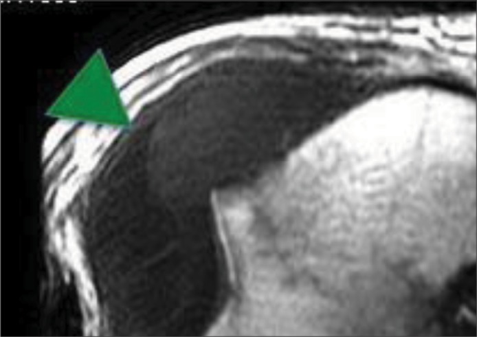
An unenhanced axial T1-weighted image at the level of the intercondylar notch demonstrates the knee joint space and synovium replaced by an intermediate soft tissue intensity mass (green arrowheads) with thin septations.

A sagittal fat-saturated T2-weighted image at the same location as the proton-density image demonstrates a mass (outlined in orange arrowheads) with associated edema, a small effusion or cystic region (yellow arrow), and distending joint space. The blue arrowhead points to a pathologic lymph node. Again, there is a minor edema signal in the tibial plateau and intercondylar notch but no aggressive subchondral osseous extension.

A sagittal proton-density image demonstrates a mass (blue arrowheads) interdigitating throughout the knee joint surrounding stretched yet intact cruciate ligaments (red arrows). There is no evidence of osseous or extraarticular extension of the mass.

A precontrast fat-saturated T1-weighted image at the level of the intercondylar notch shows persistence of the signal in the mass (orange arrowheads) on fat-saturated imaging, proving a lack of fat content and the presence of proteinaceous material.
Similar articles
-
Clinical Practice Guideline: Evaluation of the Neck Mass in Adults.Otolaryngol Head Neck Surg. 2017 Sep;157(2_suppl):S1-S30. doi: 10.1177/0194599817722550. Otolaryngol Head Neck Surg. 2017. PMID: 28891406
-
[Case report: Tenosynovial giant cell tumor arising from the knee joint].Tani Girisim Radyol. 2003 Mar;9(1):81-3. Tani Girisim Radyol. 2003. PMID: 14661299 Turkish.
-
A Case Report of Synovial Chondromatosis of the Knee Joint arising from the Marginal Synovium.J Orthop Case Rep. 2013 Jan-Mar;3(1):7-10. J Orthop Case Rep. 2013. PMID: 27298888 Free PMC article.
-
Imaging of synovial chondromatosis with radiologic-pathologic correlation.Radiographics. 2007 Sep-Oct;27(5):1465-88. doi: 10.1148/rg.275075116. Radiographics. 2007. PMID: 17848703 Review.
-
Imaging of intraarticular masses.Radiographics. 2005 Jan-Feb;25(1):105-19. doi: 10.1148/rg.251045050. Radiographics. 2005. PMID: 15653590 Review.
Cited by
-
Atypical Presentation of Metastatic Carcinoma Causing Patellar Destruction and Synovial Carcinomatosis: A Case Report.J Orthop Case Rep. 2024 Feb;14(2):59-64. doi: 10.13107/jocr.2024.v14.i02.4218. J Orthop Case Rep. 2024. PMID: 38420244 Free PMC article.
-
Image-guided synovial biopsy with a focus on neoplastic lesions.Skeletal Radiol. 2023 May;52(5):817-829. doi: 10.1007/s00256-022-04094-6. Epub 2022 Jul 22. Skeletal Radiol. 2023. PMID: 35869325 Review.
-
Synovial Metastasis From Urothelial Carcinoma of the Renal Pelvis Causing Recurrent Hemarthrosis: A Rare Presentation.Cureus. 2023 Mar 31;15(3):e36983. doi: 10.7759/cureus.36983. eCollection 2023 Mar. Cureus. 2023. PMID: 37139285 Free PMC article.
-
Calcified synovial metastasis in the knee from renal cell carcinoma: a case report.Skeletal Radiol. 2017 Jan;46(1):123-127. doi: 10.1007/s00256-016-2504-6. Epub 2016 Oct 20. Skeletal Radiol. 2017. PMID: 27761602
-
Index case of synovial metastasis in a patient with transitional cell carcinoma of the bladder.BMJ Case Rep. 2020 Jun 28;13(6):e235084. doi: 10.1136/bcr-2020-235084. BMJ Case Rep. 2020. PMID: 32595116 Free PMC article. Review.
References
-
- Thompson KS, Reyes CV, Jensen J, Gattuso P, Sacks R. Synovial metastasis: diagnosis by fine-needle aspiration cytologic investigation. Diagn Cytopathol. 1996;15(4):334–337. - PubMed
-
- Sheldon PJ, Forrester DM, Learch TJ. Imaging of intraarticular masses. Radiographics. 2005;25(1):105–119. - PubMed
-
- Ryu K, Masui F, Saito S, Marumo K. Chronic arthritis of the knee due to synovial metastasis. Jikeikai Med J. 2010;57(4):141–147.
-
- Llauger J, Palmer J, Rosón N, Bagué S, Camins A, Cremades R. Nonseptic monoarthritis: imaging features with clinical and histopathologic correlation. Radiographics. 2000;20(Spec No):S263–S278. - PubMed
LinkOut - more resources
Full Text Sources
Other Literature Sources