Comparative Study
. 2013 Jan;9(1):e1003138.
doi: 10.1371/journal.ppat.1003138. Epub 2013 Jan 31.

Novel marmoset (Callithrix jacchus) model of human Herpesvirus 6A and 6B infections: immunologic, virologic and radiologic characterization

Affiliations
Comparative Study

Novel marmoset (Callithrix jacchus) model of human Herpesvirus 6A and 6B infections: immunologic, virologic and radiologic characterization

Emily Leibovitch et al. PLoS Pathog. 2013 Jan.

Abstract

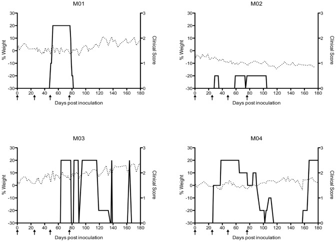
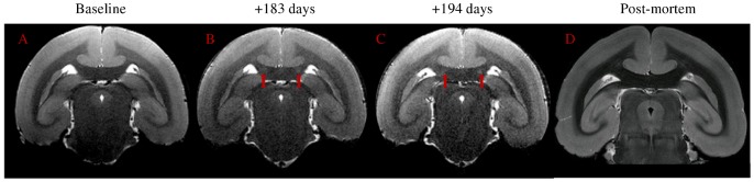
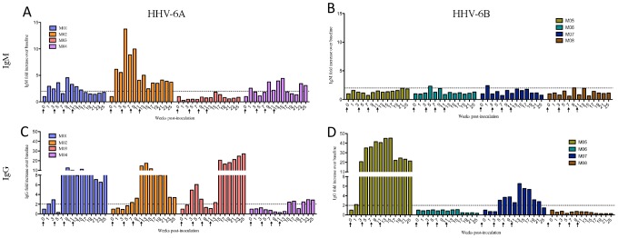
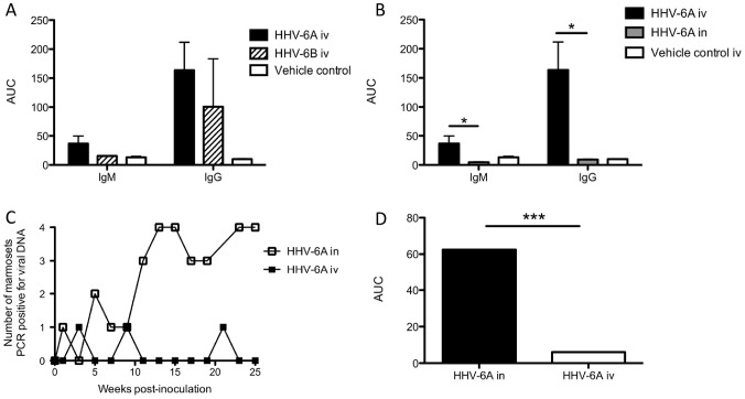
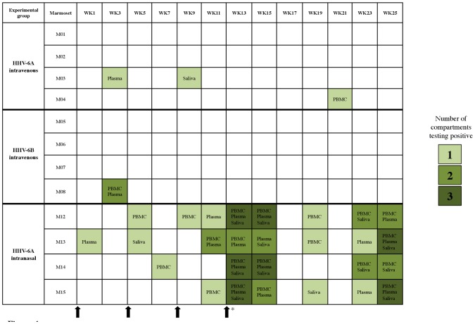
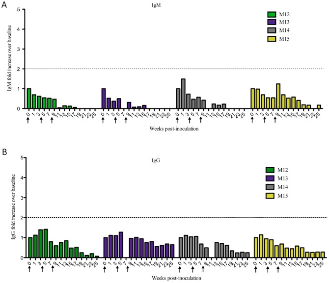
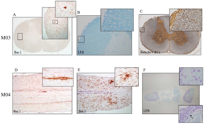
Human Herpesvirus 6 (HHV-6) is a ubiquitous virus with an estimated seroprevalence of 95% in the adult population. HHV-6 is associated with several neurologic disorders, including multiple sclerosis, an inflammatory demyelinating disease affecting the CNS. Animal models of HHV-6 infection would help clarify its role in human disease but have been slow to develop because rodents lack CD46, the receptor for cellular entry. Therefore, we investigated the effects of HHV-6 infections in a non-human primate, the common marmoset Callithrix jacchus. We inoculated a total of 12 marmosets with HHV-6A and HHV-6B intravenously and HHV-6A intranasally. Animals were monitored for 25 weeks post-inoculation clinically, immunologically and by MRI. Marmosets inoculated with HHV-6A intravenously exhibited neurologic symptoms and generated virus-specific antibody responses, while those inoculated intravenously with HHV-6B were asymptomatic and generated comparatively lower antibody responses. Viral DNA was detected at a low frequency in paraffin-embedded CNS tissue of a subset of marmosets inoculated with HHV-6A and HHV-6B intravenously. When different routes of HHV-6A inoculation were compared, intravenous inoculation resulted in virus-specific antibody responses and infrequent detection of viral DNA in the periphery, while intranasal inoculation resulted in negligible virus-specific antibody responses and frequent detection of viral DNA in the periphery. Moreover, marmosets inoculated with HHV-6A intravenously exhibited neurologic symptoms, while marmosets inoculated with HHV-6A intranasally were asymptomatic. We demonstrate that a marmoset model of HHV-6 infection can serve to further define the contribution of this ubiquitous virus to human neurologic disorders.


Conflict of interest statement

The authors have declared that no competing interests exist.

Figures

Figure 1
Figure 1. Marmosets inoculated intravenously with HHV-6A exhibited clinical symptoms without weight loss.
Percent weight change is on the left y-axis (dashed line). Clinical score is on the right y-axis (solid line). The scoring system is as follows, 0: no clinical signs, 0.5: apathy or altered walking pattern without ataxia, 1: lethargy or tremor, 2: ataxia or optic disease, 2.25: monoparesis, 2.5: paraparesis or sensory loss, 3: paraplegia or hemiplegia. Arrows represent times of HHV-6A intravenous inoculations.
Figure 2
Figure 2. Bilateral hyperintense MRI signal in corpus callosum of M04 (red arrows), inoculated with HHV-6A intravenously.
(a) Baseline, acquired before viral inoculation, (b) 183 days post-inoculation, (c) 194 days post-inoculation, (d) post-mortem scan (433 days post-inoculation).
Figure 3
Figure 3. HHV-6-specific serum antibody responses of intravenously inoculated marmosets.
Marmosets inoculated intravenously with HHV-6A generated greater virus-specific IgM and IgG responses than marmosets inoculated intravenously with HHV-6B. Plasma collected every two weeks was assayed for IgM and IgG reactivity to HHV-6 lysates. Results are represented as fold increases over baseline (before viral inoculation). The dotted line marks a two-fold increase above baseline, responses below which were considered negative. (A) IgM and (C) IgG responses of HHV-6A intravenously inoculated animals. (B) IgM and (D) IgG responses of HHV-6B intravenously inoculated animals.
Figure 4
Figure 4. Differences in HHV-6-specific antibody responses and detection of viral DNA between experimental groups.
(A) Comparison of virus-specific IgM and IgG responses between HHV-6A and HHV-6B-intravenously inoculated (iv) marmosets. AUC is calculated from Figure 3. (B) Significantly elevated virus-specific IgM and IgG responses in marmosets inoculated with HHV-6A intravenously compared to marmosets inoculated with HHV-6A intranasally (p = 0.0286, Mann Whitney U test). AUC is calculated from Figures 3, 5. (C, D) The number of marmosets testing positive for viral DNA was significantly greater in the HHV-6A intranasal group compared to the HHV-6A intravenous group (p = 0.003, Mann Whitney U test). AUC in (D) is calculated from (C). AUC: Area under the curve.
Figure 5
Figure 5. Longitudinal profiling of marmoset PBMC, plasma and saliva for HHV-6 DNA following viral inoculation.
Compartments listed in shaded boxes were positive for HHV-6 DNA by nested PCR at the indicated time points post-viral inoculation. The shading intensity corresponds to the number of positive compartments. A blank box indicates that HHV-6 DNA was not detected in PBMC, plasma or saliva. Arrows indicate times of viral inoculation, and the asterisk indicates that the fourth inoculation was specific to the HHV-6A and HHV-6B intravenous groups.
Figure 6
Figure 6. Marmosets inoculated intranasally with HHV-6A did not generate virus-specific serum IgM or IgG responses.
Plasma collected every two weeks was assayed for IgM and IgG reactivity to HHV-6 lysates. Results are represented as fold increases over baseline (before viral inoculation). The dotted line marks a two-fold increase above baseline, responses below which were considered negative. (A) IgM and (B) IgG responses of HHV-6A intranasally inoculated animals.
Figure 7
Figure 7. Spinal cord pathology in two HHV-6A intravenously inoculated marmosets.
Iba-1 is specific for microglia and macrophages, Luxol Fast Blue (LFB) stains myelin and Bielschowski's stains neurofibrils. Cervical spinal cord pathology of M03 includes (A) microglial/macrophageal aggregates identified by Iba-1, and swollen myelin sheaths identified by (B) LFB and (C) Bielschowski's. Spinal cord pathology of M04 includes microglial/macrophageal aggregates identified by Iba-1 in the (D) thoracic and (E) lumbar spinal cord and (F) myelin abnormalities identified by LFB in the dorsal root ganglia, specifically variations in sheath size and focal neuronal chromatolysis (black arrow), indicative of mild reversible damage.

References

    1. Pellett PE, Dominguez G. (2001) Chapter 80. Human Herpesviruses 6A, 6B, and 7 and Their Replication. In: Knipe DM, Howley, P.M., editor. Fields Virology. 4th edition. Philadelphia: Lippincott, Williams & Wilkins. pp. 69–2784.
    1. Salahuddin SZ, Ablashi DV, Markham PD, Josephs SF, Sturzenegger S, et al. (1986) Isolation of a new virus, HBLV, in patients with lymphoproliferative disorders. Science 234: 596–601. - PubMed
    1. Dominguez G, Dambaugh TR, Stamey FR, Dewhurst S, Inoue N, et al. (1999) Human herpesvirus 6B genome sequence: coding content and comparison with human herpesvirus 6A. J Virol 73: 8040–8052. - PMC - PubMed
    1. De Bolle L, Naesens L, De Clercq E (2005) Update on human herpesvirus 6 biology, clinical features, and therapy. Clin Microbiol Rev 18: 217–245. - PMC - PubMed
    1. De Bolle L, Van Loon J, De Clercq E, Naesens L (2005) Quantitative analysis of human herpesvirus 6 cell tropism. J Med Virol 75: 76–85. - PubMed

Publication types

MeSH terms