Review
. 2013;3(1):47-75.
doi: 10.7150/thno.5376. Epub 2013 Jan 15.

Small molecule inhibitors of CXCR4

Affiliations
Review

Small molecule inhibitors of CXCR4

Bikash Debnath et al. Theranostics. 2013.

Abstract

CXCR4 is a G-protein-coupled receptor involved in a number of physiological processes in the hematopoietic and immune systems. The SDF-1/CXCR4 axis is significantly associated with several diseases, such as HIV, cancer, WHIM syndrome, rheumatoid arthritis, pulmonary fibrosis and lupus. For example, CXCR4 is one of the major co-receptors for HIV entry into target cells, while in cancer it plays an important role in tumor cell metastasis. Several promising CXCR4 antagonists have been developed to block SDF-1/CXCR4 interactions that are currently under different stages of development. The first in class CXCR4 antagonist, plerixafor, was approved by the FDA in 2008 for the mobilization of hematopoietic stem cells and several other drugs are currently in clinical trials for cancer, HIV, and WHIM syndrome. While the long-term safety data for the first generation CXCR4 antagonists are not yet available, several new compounds are under preclinical development in an attempt to provide safer and more efficient treatment options for HIV and cancer patients.

Keywords: CXCR4; HIV; WHIM syndrome; antagonists; cancer; lupus.; rheumatoid arthritis.


Conflict of interest statement

Competing Interests: The authors have declared that no competing interest exists.

Figures

Figure 1
Figure 1
CXCR4 expression in embryonic and adult normal tissues (U133A). CXCR4 levels in various body tissues are displayed (image generated using Amazonia!: http://amazonia.transcriptome.eu 129).
Figure 2
Figure 2
CXCR4 crystal structures. A. Superimposed CXCR4 PDB structures, 3ODU (green) and 3OE0 (cyan) along with small molecule ligand IT1t (pink) and peptidic ligand CVX15 (yellow); B. Binding site of CXCR4 - a small conformational changes are visible between those two structures in the binding site region. IT1t (pink); CVX15 (yellow); binding site residues from 3ODU (green) and 3OE0 (cyan).
Figure 3
Figure 3
CXCR4 mediates HIV infection and cancer progression. CXCR4 is a co-receptor used along with CD4 by HIV-1 strains for infecting T cells. The binding of gp120 to CD4 induces a conformational change of gp120, allowing it to interact with CXCR4's N-terminal, ECL2 and ECL3 domains as well as the ligand binding cavity through the V3 loop of gp120. These interactions trigger a conformational change in gp41, causing a pH-dependent fusion of the viral and the host cell membranes and thus the delivery of the viral payload. CXCR4 is also involved in the progression of tumor (hematopoietic and solid) via the interaction with SDF-1. The SDF-1/CXCR4 interaction stimulates tyrosine phosphorylation of CXCR4, followed by the activation of multiple G protein-dependent signaling pathways, which may be different among cell types. The major potential pathways and their cellular outcomes are described in this figure. Activated CXCR4 recruits arrestin 2/3, which mediate receptor internalization. CXCR4 may then be recycled to the cell surface or ubiquitinated by AIP4 that results in the lysosomal degradation of CXCR4.
Figure 4
Figure 4
Structure of select CXCR4 antagonists , .
Figure 5
Figure 5
Structures of select cyclic pentapeptide-based CXCR4 antagonists. IC50 values are the concentrations for 50% inhibition of the [125I]-SDF- 1α binding to CXCR4 transfectant of HEK293 cells .
Figure 6
Figure 6
Structures of representative examples of cyclic pentapeptide-based CXCR4 antagonists .
Figure 7
Figure 7
Structures of representative examples of a monomeric cyclic pentapeptide-based CXCR4 antagonist, two dimeric derivatives and a dimeric Ga-labeled DOTA complex .
Figure 8
Figure 8
Structures of indole-based CXCR4 antagonists .
Figure 9
Figure 9
General structure (48) and representative examples of tetrahydroquinoline-based CXCR4 antagonists. IC50 values were determined in antiviral assay using HOS cells expressing hCXCR4/hCCR5/hCD4/pHIV-LTR-luciferase and HIV-1 IIIB strain .
Figure 10
Figure 10
Representative examples of tetrahydroquinoline-based CXCR4 antagonists. IC50 values were determined in antiviral assay using HOS cells expressing hCXCR4/hCCR5/hCD4/pHIV-LTR-luciferase and HIV-1 IIIB strain .
Figure 11
Figure 11
Select examples of tetrahyidroquinoline-based CXCR4 antagonists. IC50 values were determined in antiviral assay using HOS cells expressing hCXCR4/hCCR5/hCD4/pHIV-LTR-luciferase and HIV-1 IIIB strain .
Figure 12
Figure 12
Select examples of tetrahydroquinoline-based CXCR4 antagonists. IC50 values were determined in antiviral assay using HOS cells expressing hCXCR4/hCCR5/hCD4/pHIV-LTR-luciferase and HIV-1 IIIB strain .
Figure 13
Figure 13
Representative examples of tetrahydroquinoline-based CXCR4 antagonists. IC50 values are the concentrations required to protect 50% of MT4 cells against X4 HIV-1 NL4.3 cytopathicity .
Figure 14
Figure 14
Select examples of tetrahydroquinoline-based CXCR4 antagonists. IC50 values are the concentrations required to protect 50% of MT4 cells (compounds 105-114) or HOS cells (compounds 115, 116) against viral cytopathicity , .
Figure 15
Figure 15
Select examples of tetrahydroquinoline-based CXCR4 antagonists. IC50 values were determined in antiviral assay using HOS cells expressing hCXCR4/hCCR5/hCD4/pHIV-LTR-luciferase and HIV-1 IIIB strain .
Figure 16
Figure 16
Select examples of tetrahydroquinoline-based CXCR4 antagonists. IC50 values are the concentrations required to protect 50% MT4 cells against X4 HIV-1 NL4.3 cytopathicity .
Figure 17
Figure 17
Representative examples of p-xylyl-enediamine based CXCR4 antagonists. EC50 values are the concentrations required to protect 50% of HIV-1 IIIB strain infected MT-4 cells against viral cytopathicity .
Figure 18
Figure 18
Select examples of p-xylyl-enediamine based CXCR4 antagonists. EC50 values are the concentrations required to protect 50% of HIV-1 IIIB strain infected MT-4 cells against viral cytopathicity .
Figure 19
Figure 19
Select examples of p-xylyl-enediamine based CXCR4 antagonists. EC (effective concentration) values are the concentrations at which the compound blocks > 50% of the peptide TN14003 binding to CXCR4 .
Figure 20
Figure 20
Starting amines for preparation of guanide, biguanide, or phenylguanide derivatives .
Figure 21
Figure 21
Structure of quinoline based CXCR4 antagonists. EC50 values are the concentrations for 50% inhibition of the SDF-1α binding to CXCR4 receptor in HEK293 cells .
Figure 22
Figure 22
Representative examples of pyrimidine-based CXCR4 antagonists .
Figure 23
Figure 23
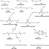
Miscellaneous structures of CXCR4 antagonists -.

References

    1. Lefrancois M, Lefebvre MR, Saint-Onge G, Boulais PE, Lamothe S, Leduc R. et al. Agonists for the Chemokine Receptor CXCR4. ACS Med Chem Lett. 2011;2:597–602. - PMC - PubMed
    1. Zhan W, Liang Z, Zhu A, Kurtkaya S, Shim H, Snyder JP. et al. Discovery of small molecule CXCR4 antagonists. J Med Chem. 2007;50:5655–64. - PubMed
    1. Bleul CC, Wu L, Hoxie JA, Springer TA, Mackay CR. The HIV coreceptors CXCR4 and CCR5 are differentially expressed and regulated on human T lymphocytes. Proc Natl Acad Sci U S A. 1997;94:1925–30. - PMC - PubMed
    1. Oberlin E, Amara A, Bachelerie F, Bessia C, Virelizier JL, Arenzana-Seisdedos F. et al. The CXC chemokine SDF-1 is the ligand for LESTR/fusin and prevents infection by T-cell-line-adapted HIV-1. Nature. 1996;382:833–5. - PubMed
    1. Bleul CC, Farzan M, Choe H, Parolin C, Clark-Lewis I, Sodroski J. et al. The lymphocyte chemoattractant SDF-1 is a ligand for LESTR/fusin and blocks HIV-1 entry. Nature. 1996;382:829–33. - PubMed