. 2013;8(1):e48814.
doi: 10.1371/journal.pone.0048814. Epub 2013 Jan 30.

Differential expression of exosomal microRNAs in prefrontal cortices of schizophrenia and bipolar disorder patients

Affiliations

Differential expression of exosomal microRNAs in prefrontal cortices of schizophrenia and bipolar disorder patients

Meredith G Banigan et al. PLoS One. 2013.

Abstract

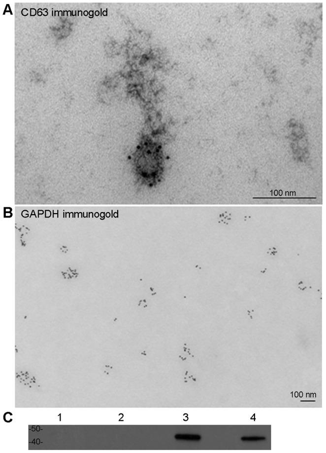
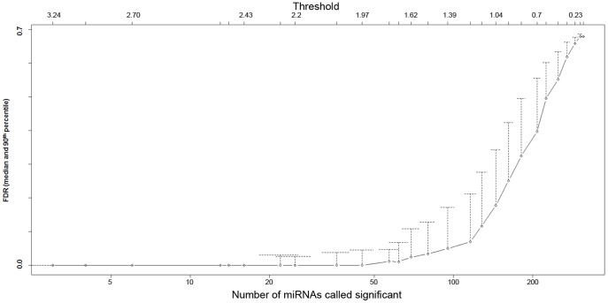
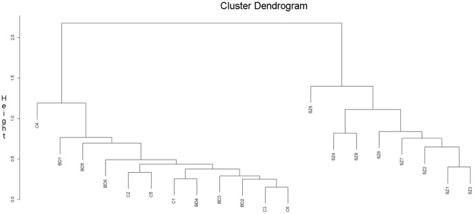
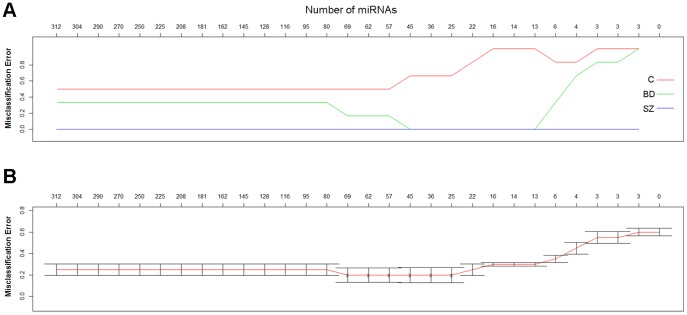
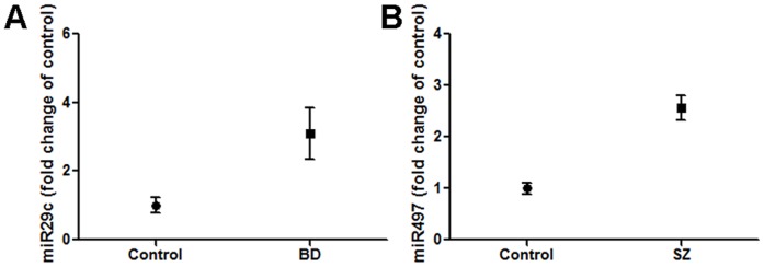
Exosomes are cellular secretory vesicles containing microRNAs (miRNAs). Once secreted, exosomes are able to attach to recipient cells and release miRNAs potentially modulating the function of the recipient cell. We hypothesized that exosomal miRNA expression in brains of patients diagnosed with schizophrenia (SZ) and bipolar disorder (BD) might differ from controls, reflecting either disease-specific or common aberrations in SZ and BD patients. The sources of the analyzed samples included McLean 66 Cohort Collection (Harvard Brain Tissue Resource Center), BrainNet Europe II (BNE, a consortium of 18 brain banks across Europe) and Boston Medical Center (BMC). Exosomal miRNAs from frozen postmortem prefrontal cortices with well-preserved RNA were isolated and submitted to profiling by Luminex FLEXMAP 3D microfluidic device. Multiple statistical analyses of microarray data suggested that certain exosomal miRNAs were differentially expressed in SZ and BD subjects in comparison to controls. RT-PCR validation confirmed that two miRNAs, miR-497 in SZ samples and miR-29c in BD samples, have significantly increased expression when compared to control samples. These results warrant future studies to evaluate the potential of exosome-derived miRNAs to serve as biomarkers of SZ and BD.


Conflict of interest statement

Competing Interests: The authors have declared that no competing interests exist.

Figures

Figure 1
Figure 1. Characterization of exosome-containing pellets from human brain tissue.
Electron microscopy of exosomal extractions from BA9 cortices demonstrates the presence of microvesicles (∼70–100 nm in diameter). Upon immuno-gold labeling procedure with antibodies against CD63 (A; additional negative staining highlights membrane of the vesicle) and GAPDH (B), the microvesicles reveal the presence of exosome-associated antigens. Bars indicate 100 nm. Comparison of exosomal extraction procedure products from BA9 cortices and H4 cell-culture reveals similar outcomes in Western blot. Supernatant of BA9 exosome-containing pellets (lane 1), supernatant of H4 exosome-containing pellets (lane 2), BA9 exosome-containing pellets reconstituted in PBS (lane 3), and of H4 exosome-containing pellets reconstituted in PBS (lane 4), show robust presence of exosomal marker flotillin-2 in the pellets, but not in the supernatants (C).
Figure 2
Figure 2. Median and 90th percentile of False Discovery Rates (FDR) in order left-to-right of threshold value and number of statistically significant results for all three groups (C, BD, and SZ).
The Y-axis is an estimate of the percentage of false positives. High-ranked miRNA (at the left) have a low rate of false positives, while lower-ranked miRNA (moving toward the right) have higher rates of false positives.
Figure 3
Figure 3. Hierarchical clustering analysis of top 21 ranked miRNAs from FDR analysis.
Correlation coefficient (cc) was generated to assess the relationship between the expression values of each sample and the rest of the samples (see Methods). The coefficient is 1 if their expression profiles are highly similar and 0 if their expression profiles are highly divergent. The clustering graph is built so that the samples with similar expression patterns are clustered at the bottom while more differential patterns are at the top of the graph.
Figure 4
Figure 4. Misclassification rate analysis.
(A) Controls (red) have the most variable miRNA expression. The expression data have less variability in BD (green) and the least in SZ (blue; misclassification rate  = 0), resulting in stronger predictive power within their respective clinical groups.
Figure 5
Figure 5. In comparison to controls, the expression of miR-29c and miR-497 is significantly increased in BD and SZ samples, respectively.
Average exosomal RNA extracted from BA9 cortices of BD samples show a 2.77 fold increase of miR-29c in comparison to controls (A). SZ samples reveal 2.35 fold increase of miR-497 when compared to controls (B). Error bars indicate S.E.M.

References

    1. De Smaele E, Ferretti E, Gulino A (2010) MicroRNAs as biomarkers for CNS cancer and other disorders. Brain Res 1338: 100–111. - PubMed
    1. Beveridge NJ, Cairns MJ (2011) MicroRNA dysregulation in schizophrenia. Neurobiol Dis 46: 263–71. - PubMed
    1. Sempere LF, Freemantle S, Pitha-Rowe I, Moss E, Dmitrovsky E, et al. (2004) Expression profiling of mammalian microRNAs uncovers a subset of brain-expressed microRNAs with possible roles in murine and human neuronal differentiation. Genome Biol 5: R13. - PMC - PubMed
    1. Ambros V (2003) MicroRNA pathways in flies and worms: growth, death, fat, stress, and timing. Cell 113: 673–676. - PubMed
    1. Kim J, Inoue K, Ishii J, Vanti WB, Voronov SV, et al. (2007) A MicroRNA feedback circuit in midbrain dopamine neurons. Science 317: 1220–1224. - PMC - PubMed

Publication types