. 2013;8(1):e50825.
doi: 10.1371/journal.pone.0050825. Epub 2013 Jan 31.

The phosphoinositide-binding protein ZF21 regulates ECM degradation by invadopodia

Affiliations

The phosphoinositide-binding protein ZF21 regulates ECM degradation by invadopodia

Daisuke Hoshino et al. PLoS One. 2013.

Abstract

During the process of tumor invasion, cells require footholds on extracellular matrices (ECM) that are created by forming focal adhesions (FAs) using integrins. On the other hand, cells must degrade the ECM barrier using extracellular proteases including MMPs in the direction of cell movement. Degradation occurs at the leading edges or invadopodia of cells, which are enriched in proteases and adhesion molecules. Recently, we showed that the phosphoinositide-binding protein ZF21 regulates FA disassembly. ZF21 increased cell migration by promoting the turnover of FAs. In addition, ZF21 promotes experimental tumor metastasis to lung in mice and its depletion suppresses it. However, it is not known whether ZF21 regulates cancer cell invasion in addition to its activity on FAs. In this study, we demonstrate that ZF21 also regulates invasion of tumor cells, whereas it does not affect the overall production of MMP-2, MMP-9, and MT1-MMP by the cells. Also, we observe that the ECM-degrading activity specifically at the invadopodia is severely abrogated. In the ZF21 depleted cells MT1-MMP cannot accumulate to the invadopodia and thereby cannot contribute to the ECM degradation. Thus, this study demonstrates that ZF21 is a key player regulating multiple aspects of cancer cell migration and invasion. Possible mechanisms regulating ECM degradation at the invadopodia are discussed.


Conflict of interest statement

Competing Interests: The authors have declared that no competing interests exist.

Figures

Figure 1
Figure 1. ZF21-knockdown increases the number of FAs and reduces the invasive activity of HT1080 cells.
A. Expression of ZF21 in HT1080 cells was knocked down using either of two shRNA sequences targeting ZF21 mRNA (shZF21#1 and #2). Endogenous ZF21 was detected by Western blot analysis by using a polyclonal anti-ZF21 antibody. B. The cells expressing shLacZ (top) or shZF21#1 and #2 (middle and bottom) were seeded onto glass coverslips. After 48 h, FAK phosphorylated at Tyr397 (pY397-FAK) was visualized with a specific antibody. Scale bar, 10 µm. C. Quantitative analysis of the number of pY397-FAK positive punctate signals. The number of pY397-FAK positive punctate singals was counted in 100 cells. The experiment was independently repeated three times. D. The cells expressing shLacZ or shZF21#1 and #2 were subjected to a migration (a) and a matrigel invasion assay (b) using a transwell chamber equipped with filters coated with fibronectin or matrigel. As an attractant, fetal bovine serum was added in lower chamber. Error bars indicate the means±S.D. (n = 3).*, p<0.05 (Student's t test).
Figure 2
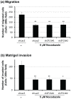
Figure 2. Microtubule disruption diminishes the effect of ZF21-knockdown on the invasive activity of the cells.
In the presence of 5 µM nocodazole, the same set of cells indicated in Fig. 1D was subjected to migration (a) and matrigel invasion assays (b). Error bars indicate the means±S.D. (n = 3).*, p<0.05 **, p<0.01 (Student's t test).
Figure 3
Figure 3. ZF21-mediated matrigel invasion was suppressed by MMP inhibition.
A. In the presence of 10 µM MMI270, the same set of cells indicated in Fig. 1D was subjected to migration (a) and matrigel invasion assays (b). Error bars indicate the means±S.D. (n = 3).*, p<0.01 **, p<0.005 (Student's t test). B. The cultured media of HT1080 cells expressing shLacZ or shZF21#1 and #2 were subjected to gelatin zymography analysis to detect MMP-2 and MMP-9. The same samples indicated in Fig. 1A were subjected to the Western blot analysis to detect MT1-MMP using a specific antibody. The expression levels of MT1-MMP in the cells were normalized by those of actin indicated in Fig. 1A.
Figure 4
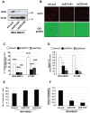
Figure 4. ZF21 knockdown reduces invadopodia-mediated ECM degradation.
A. Expression of ZF21 in MDA-MB231 cells was knocked down using either of two shRNA sequences targeting ZF21 mRNA (shZF21#1 and #2). Endogenous ZF21 was detected by Western blot analysis by using a polyclonal anti-ZF21 antibody. B. The cells expressing shLacZ (top) or shZF21#1 and #2 (middle and bottom) cultured on Oregon Green-labeled gelatin-coated glass coverslips. After fixation, the cells were stained with phalloidin for localization of F-actin (red on left panels). Degraded ECM appears as dark areas (reduced green fluorescence on right panels). (scale bar, 10 μm). C. Percentage of cells exhibiting ECM degradation. Number of cells stained with phalloidin was counted and merged with the ECM degraded area. The averaged number of the ECM-degrading cells and total counted cells are indicated upon each column in graphs (ECM-degrading cells/total counted cells). Ratio of the ECM-degrading cells is represent as the means ± S.D (n = 3), *, P<0.0001, **, P<0.005. D. Average degradation area per cell is calculated and presented. Error bars indicate the means ± S.E.M. *, P<0.0001, **, P<0.005. E. Number of punctate actin signals was counted for 450–500 cells. Average number of actin dots per cell is presented. Error bars indicate the means ± S.E.M *, P<0.0005. The experiment was repeated six times. F. Ratio of ECM-degrading actin dots (active invadopodia) to no ECM-degrading actin dots (inactive invadopodia) is presented. Error bars indicate the means ± S.E.M *, P<0.0005.
Figure 5
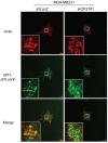
Figure 5. ZF21 regulates localization of MT1-MMP to invadopodia.
MT1-MMP-pHLuorin (MT1-pHLuorin) was transiently expressed in the MDA-MB231 cells expressing either shLacZ or shZF21#1. The cells were cultured on cover slips coated with 50 µg/ml fibronectin for 6 hours. After fixation, the cells were stained with Rhodamine-labeled phalloidin to visualize F-actin. The localizations of F-actin (red on top panels) or MT1-MMP-pHLuorin (green on middle panels) were observed using confocal microscopy. The merged pictures of red and green fluorescence were shown on bottom panels. The boxed areas are shown at higher magnification in the panels. (scale bar, 10 μm).

Similar articles

Cited by

References

    1. Gupta GP, Massague J (2006) Cancer metastasis: building a framework. Cell 127: 679–695. - PubMed
    1. Steeg PS (2006) Tumor metastasis: mechanistic insights and clinical challenges. Nat Med 12: 895–904. - PubMed
    1. Guo W, Giancotti FG (2004) Integrin signalling during tumour progression. Nat Rev Mol Cell Biol 5: 816–826. - PubMed
    1. Parsons JT, Horwitz AR, Schwartz MA (2010) Cell adhesion: integrating cytoskeletal dynamics and cellular tension. Nat Rev Mol Cell Biol 11: 633–643. - PMC - PubMed
    1. Petit V, Thiery JP (2000) Focal adhesions: structure and dynamics. Biol Cell 92: 477–494. - PubMed

Publication types

Substances