. 2013;8(1):e51573.
doi: 10.1371/journal.pone.0051573. Epub 2013 Jan 28.

The mechanism of effective electroacupuncture on T cell response in rats with experimental autoimmune encephalomyelitis

Affiliations

The mechanism of effective electroacupuncture on T cell response in rats with experimental autoimmune encephalomyelitis

Yumei Liu et al. PLoS One. 2013.

Abstract

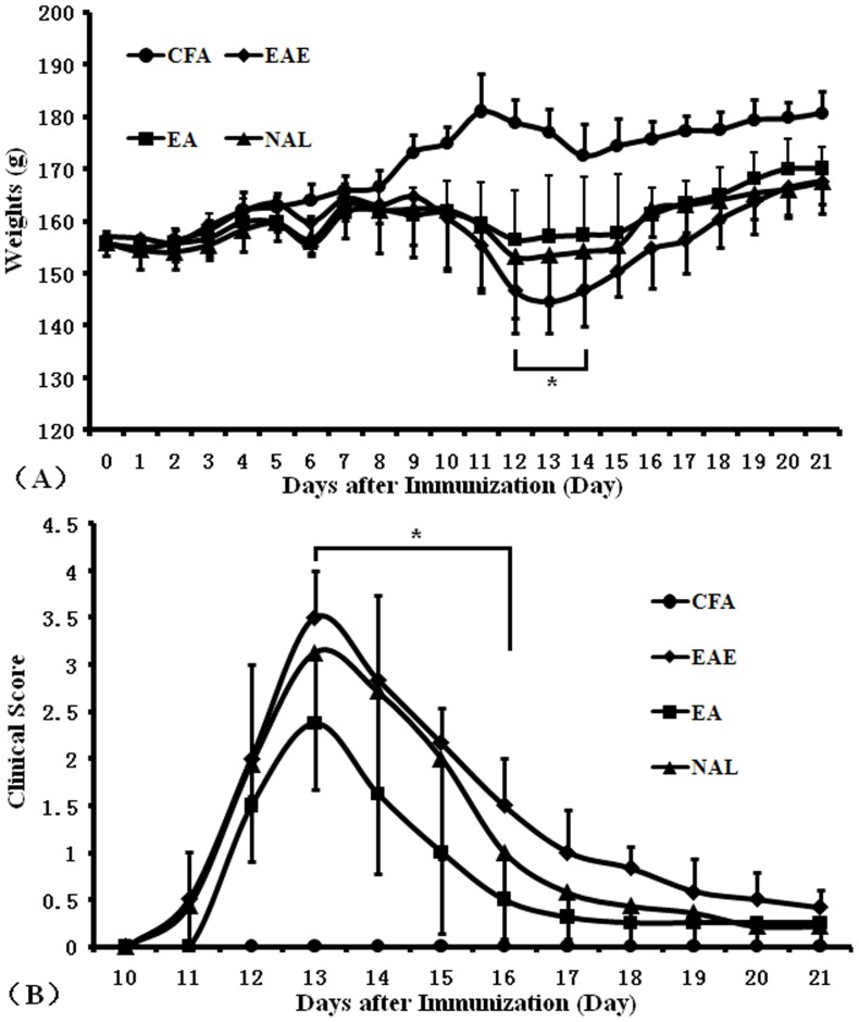
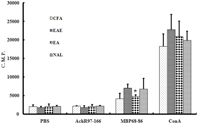
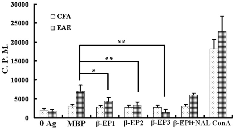
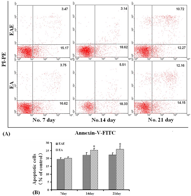
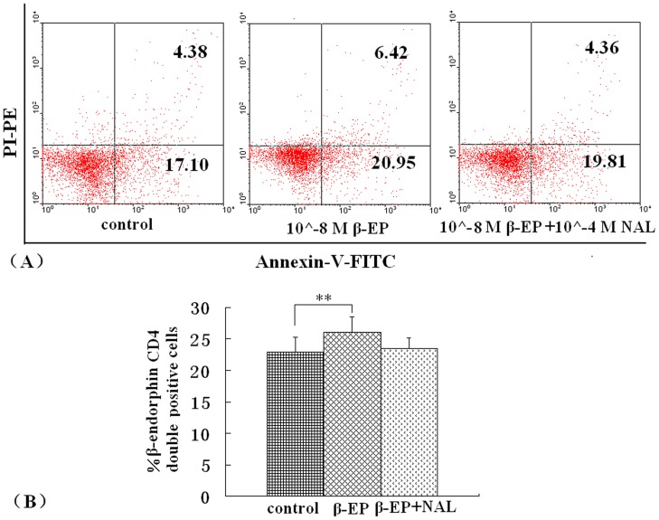
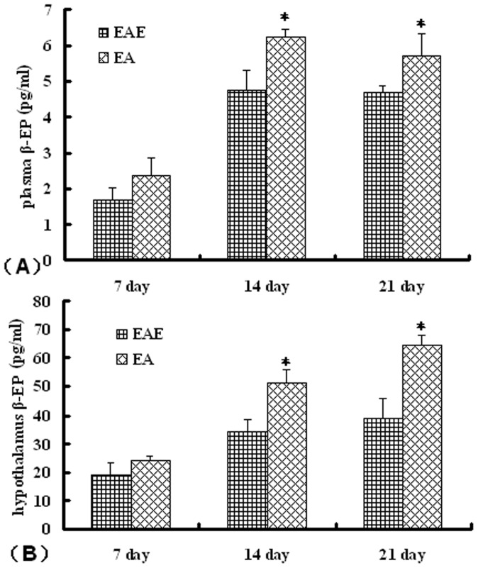
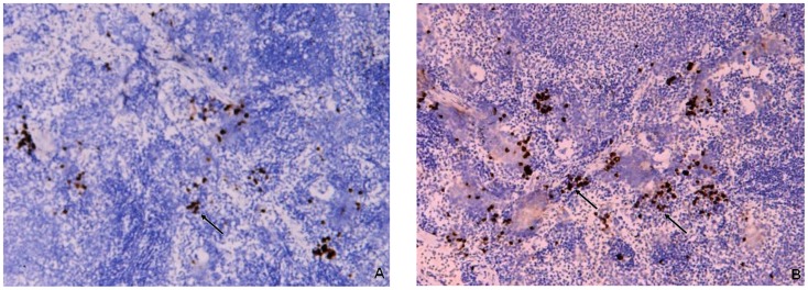
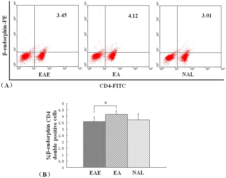
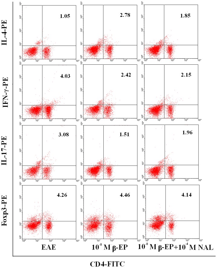
Previously, we demonstrated that electroacupuncture (EA) decreased lymphocyte infiltration into the spinal cords of rats presenting with experimental autoimmune encephalomyelitis (EAE), a disease model used in the study of multiple sclerosis (MS). The aim of this study was to characterize the effects of EA on the EAE. Female Lewis rats were divided into either CFA, EAE, EA, or injection with naloxone after electroacupuncture (NAL) groups. Electroacupuncture was administered every day for 21 days. To evaluate proliferation and apoptosis, lymphocytes from rats presenting with EAE were collected and cultured with β-endorphin. Immunohistochemisty, flow cytometry and radio-immunity methods were applied to detect the expression of β-endorphin. Results presented in this report demonstrate that the beneficial anti-inflammatory effects of EA on EAE were related to β-endorphin production that balances the Thl/Th2 and Th17/Treg responses. These results suggest that β-endorphin could be an important component in the development of EA-based therapies used for the treatment of EAE.


Conflict of interest statement

Competing Interests: The authors have declared that no competing interests exist.

Figures

Figure 1
Figure 1. Body weight and clinical scores.
After the first immunization, mice were weighed and disease severity scored daily until day 21 post immunization. Body weights (A) and clinical scores (B) were measured in control rats (•), EAE rats (♦), EA rats (▪), and NAL rats (▴) (n = 8 for EAE and NAL groups, n = 9 for EA groups, n = 5 for the CFA group, *P<0.05). The results shown are representative of 3 separate experiments.
Figure 2
Figure 2. The effect of EA treatment on lymphocyte proliferation.
Lymphocytes were isolated from CFA, EAE, EA, and, NAL rats 14 days post immunization. Cells were incubated with or without MBP68–86 (10 mg/ml) or AchR97–166 (10 µg/ml) or ConA (5 mg/ml) for 48 h. Cell proliferation was assessed by [3H]thymidine incorporation. Proliferation of lymphocytes from EA-treated rats was reduced. The results are shown as mean counts per minute (C.P.M.) ± SE. *P<0.05 vs. EAE group.
Figure 3
Figure 3. β-endorphin inhibits antigen-dependent proliferation of lymphocytes from EAE rats.
Proliferation of rat CD4+ MBP specific T cells or CD4+ non-specific T cells stimulated with or without antigen in the absence or presence of different concentrations of β-endorphin and/or naloxone was assessed. β-EP1∶10−8 M β-endorphin, β-EP2∶10−7 M β-endorphin, β-EP3∶10−6 M β-endorphin, β-EP1+NAL: 10−8 M β-endorphin+10−4 M naloxone, *P<0.05, **P<0.01.
Figure 4
Figure 4. Apoptosis measurements.
Apoptosis was determined by flow cytometric analysis using double staining of cells with Annexin V/PI. (A). Representative flow cytometric analysis of cells harvested from rats in the EAE and EA groups. (B). Percent number of cells undergoing apoptosis in rats from the EAE and EA groups over time. *P<0.05.
Figure 5
Figure 5. Effect of β-endorphin on lymphocytes apoptosis.
Lymphocytes were harvested from EAE and cultured with 10−8 M β-endorphin. To detect apoptotic lymphocytes flow cytometric analysis was applied. (A). Representative flow cytometric analysis of apoptotic cells. (B). Percent number of cells undergoing apoptosis in the EAE lymphocytes, cultured with β-endorphin or β-endorphin and nalxone. **P<0.01 control vs. 10−8 M β-endorphin.
Figure 6
Figure 6. Effect of EA on β-endorphin levels in EAE rats.
Rats from the respective groups were sacrificed at 7, 14, or 21 d post-immunization with MBP68–86 and the β-endorphin levels present in (A) plasma and (B) hypothalamus determined. Values are expressed as the mean ± SE of x observations (n = 5, *P<0.05, **P<0.01 EA vs. EAE group).
Figure 7
Figure 7. Expression of β-endorphin in spleen tissue sections.
Immunohistochemical staining of β-endorphin positive cells in rat splenic tissues from EAE and EA rats 14 days post immunization. (A) β-endorphin positive cells in EAE rats. (B) β-endorphin positive cells in EA rats. Magnification: ×100.
Figure 8
Figure 8. Effect of EA on CD4+ T cell β-endorphin expression in EAE rats.
Lymphocytes from either EAE, EA, or NAL rat groups were isolated on day 14 post immunization. CD4+ cell expression of β-endorphin was assessed by flow cytometry. (A). Representative flow cytometric analysis of cells harvested from rats in the EAE, EA and NAL groups. (B). Percent number of CD4+ T cells β-endorphin expression in rats from the EAE, EA and NAL groups. *P<0.05.
Figure 9
Figure 9. Subtype changes in MBP68–86-specific lymphocytes following co-culture with β-endorphin and/or naloxone.
Lymphocytes co-cultured with β-endorphin and/or naloxone for 72 h cells were collected and CD4, IFN-γ, IL-4, IL-17, and FoxP3 expression levels analyzed by flow cytometry. Representative results from 3 separate experiments are shown.

Similar articles

Cited by

References

    1. Gironi M, Martinelli V, Brambilla E, Furlan R, Panerai AE, et al. (2000) Beta-endorphin concentrations in peripheral blood mononuclear cells of patients with multiple sclerosis: effects of treatment with interferon beta. Arch Neurol 57: 1178–1181. - PubMed
    1. Yoshino S, Koiwa M, Shiga H, Nakamura H, Higaki M, et al. (1992) Miyasaka N. Detection of opioid peptides in synovial tissues of patients with rheumatoid arthritis. J Rhematol 19: 660–661. - PubMed
    1. Börner C, Warnick B, Smida M, Hartig R, Lindquist JA, et al. (2009) Mechanisms of opioid-mediated inhibition of human T cell receptor signaling. J Immunol. 183(2): 882–889. - PubMed
    1. Crine P, Gianoulakis C, Seidah NG (1978) Biosynthesis of beta-endorphin from beta-lipotropin and a larger molecular weight precursor in rat pars intermedia. Proc Natl Acad Sci U S A 75: 4719–4723. - PMC - PubMed
    1. Panerai AE, Sacerdote P (1997) Beta-endorphin in the immune system: a role at last? Immunol Today 18: 317–319. - PubMed

Publication types

MeSH terms