. 2013;8(1):e54267.
doi: 10.1371/journal.pone.0054267. Epub 2013 Jan 30.

Blockade of Kv1.3 potassium channels inhibits differentiation and granzyme B secretion of human CD8+ T effector memory lymphocytes

Affiliations

Blockade of Kv1.3 potassium channels inhibits differentiation and granzyme B secretion of human CD8+ T effector memory lymphocytes

Lina Hu et al. PLoS One. 2013.

Abstract

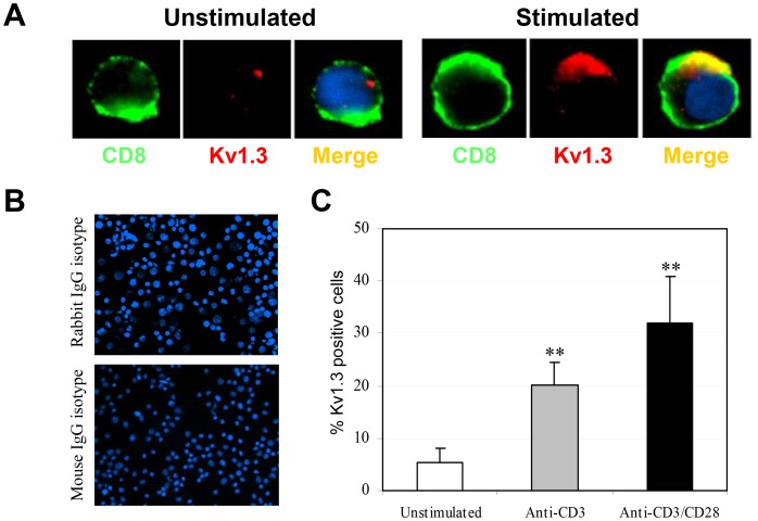
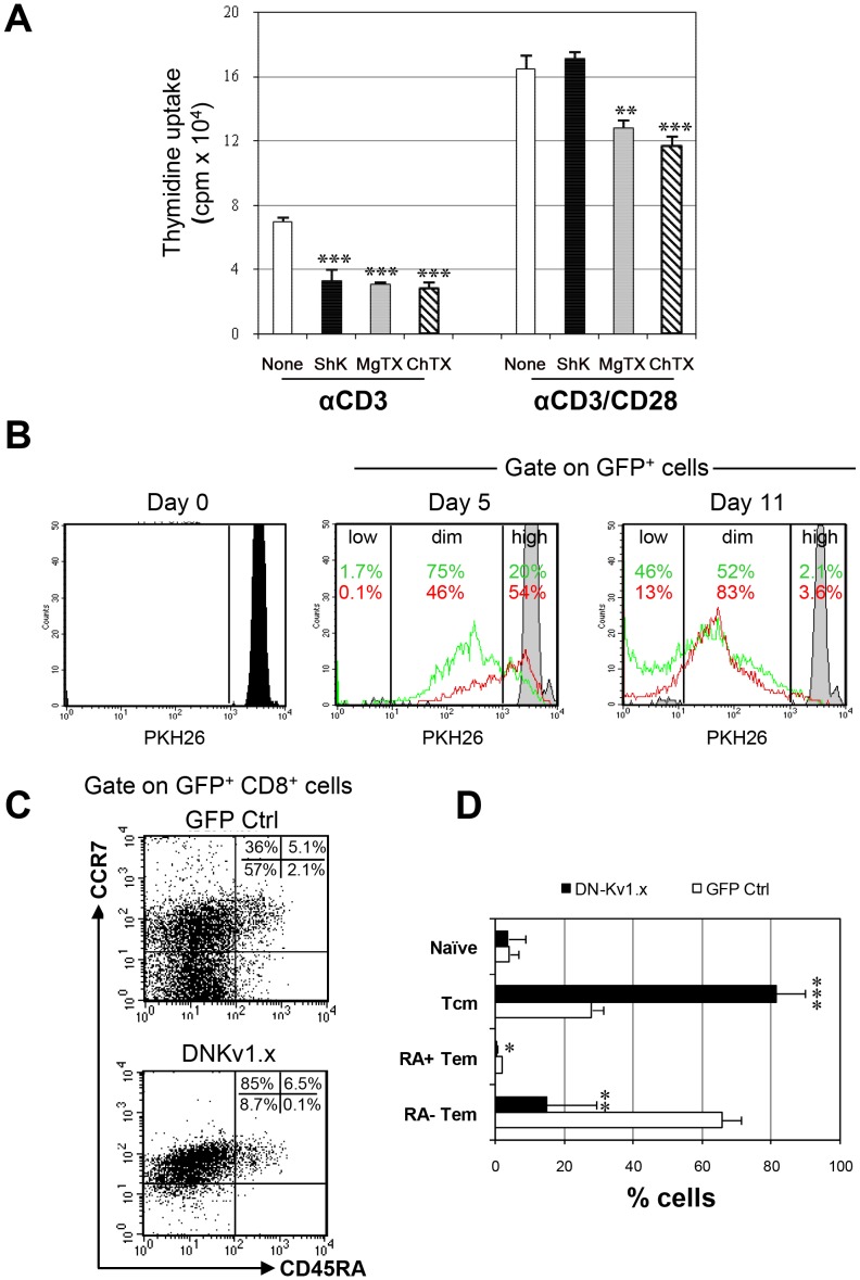
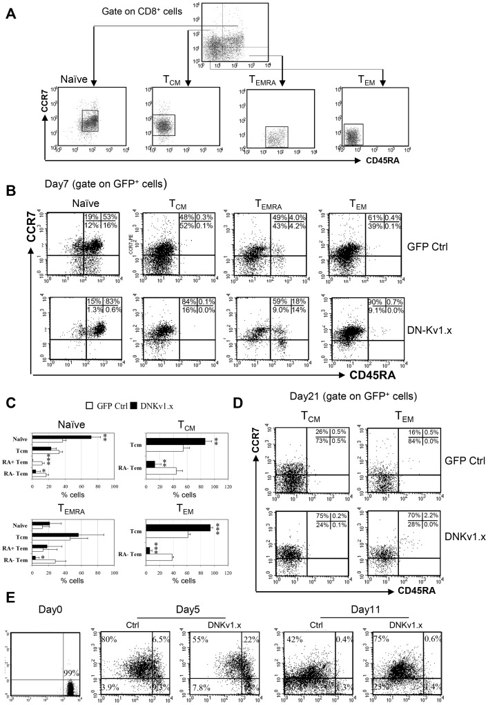
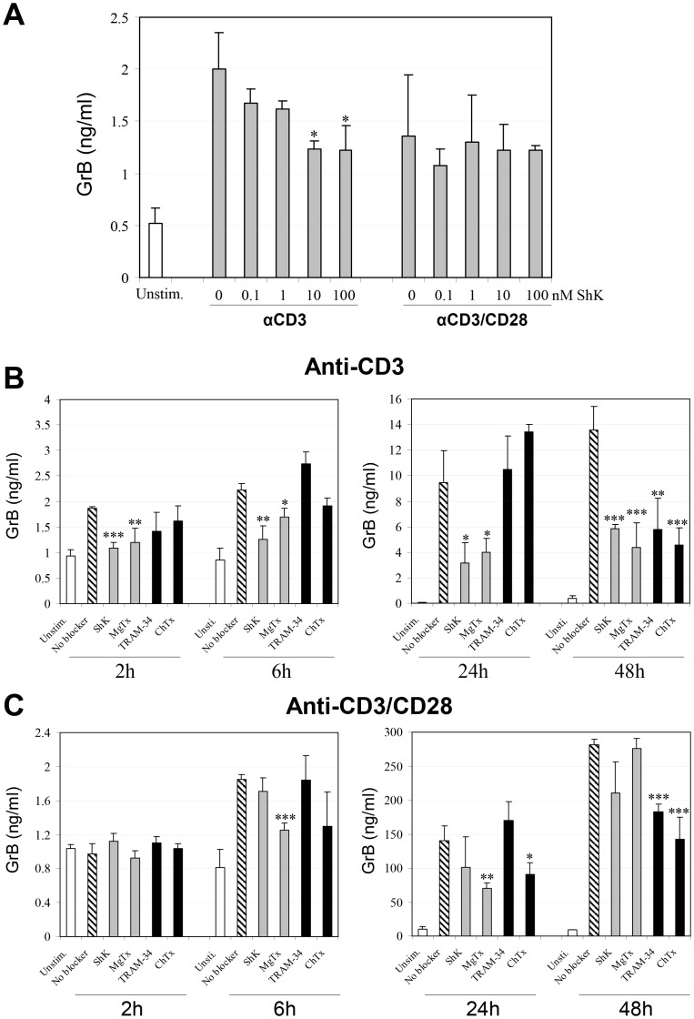
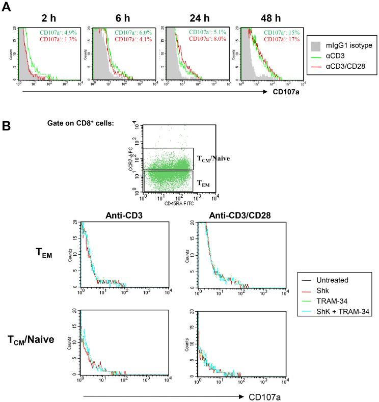
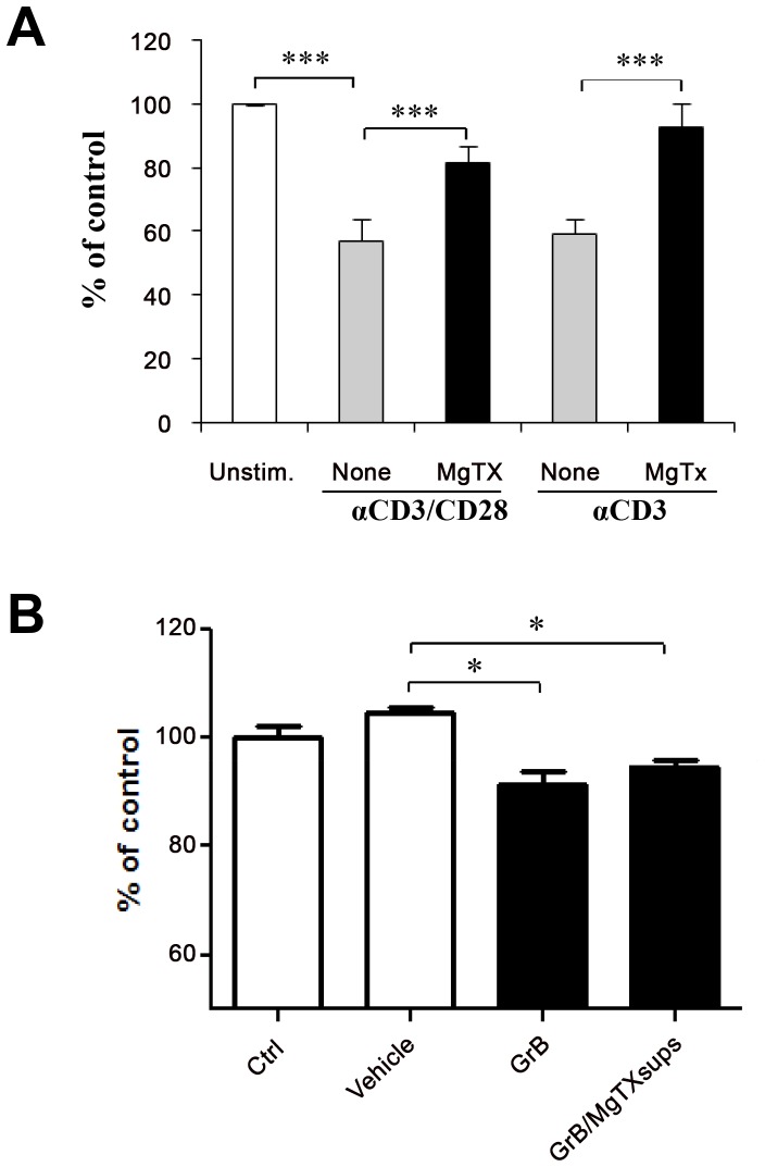
Increased expression of the voltage-gated potassium channel Kν1.3 on activated effector memory T cells (T(EM)) is associated with pathology in multiple sclerosis (MS). To date, most studies of Kν1.3 channels in MS have focused on CD4+ T(EM) cells. Much less is known about the functional relevance of Kv1.3 on CD8+ T(EM) cells. Herein, we examined the effects of Kν1.3 blockade on CD8+ T cell proliferation, differentiation into cytotoxic effector cells, and release of granzyme B (GrB), a key effector of CD8+ T cell-mediated cytotoxicity. We confirmed the expression of Kv1.3 channels on activated human CD8+ T lymphocytes by immunofluorescent staining. To test the functional relevance of the Kv1.3 channel in CD8+ T cells, we inhibited this channel via pharmacological blockers or a lentiviral-dominant negative (Kv1.xDN) approach and determined the effects of the blockade on critical pathogenic parameters of CD8+ T cells. We found that blockade of Kv1.3 with both lentivirus and pharmacologic agents effectively inhibited cytotoxic effector memory cells' proliferation, secretion of GrB, and their ability to kill neural progenitor cells. Intriguingly, the KvDN transduced T cells exhibited arrested differentiation from central memory (T(CM)) to effector memory (T(EM)) states. Transduction of cells that had already differentiated into T(EM) with KvDN led to their conversion into T(CM). CD8+ T(EM) have a critical role in MS and other autoimmune diseases. Our present results indicate a critical role for Kv1.3 in the conversion of CD8+ T cells into potential pathogenic effector cells with cytotoxic function.


Conflict of interest statement

Competing Interests: The authors have declared that no competing interests exist.

Figures

Figure 1
Figure 1. Kv1.3 expression is upregulated in activated CD8+ T cells and co-localizes with CD8.
(A) Purified CD8+ T cells were stimulated with anti-CD3/CD28 for 3 days. Naïve and stimulated cells were then immunostained for Kv1.3 in combination with CD8 and subsequently viewed by immunofluorescence microscopy. Cellular nuclei were counterstained with DNA dye DAPI (blue). Kv1.3 detected by AF 594 fluorescence is shown in red, while CD8 detected by AF 488 fluorescence is shown in green. Colocalization is indicated by a yellow and/or orange color in the overlay panels. (B) An isotype-matched antibody was used as a negative control. Original magnification, ×100. Image is representative of three different donors. (C) Summary of percentages of activated CD8+ T cells expressing Kv1.3. In brief, 4 view fields/microscopic section were evaluated for Kv1.3+ CD8 cells stimulated with anti-CD3/CD28 or anti-CD3 alone for 3 days. The percentages of Kv1.3+ cells are based on the number of CD8+ T cells counted. Data are mean ± SD from one representative of three independent and reproducible experiments. Values that are significantly different from that of non-stimulated control are indicated as **, p<0.01.
Figure 2
Figure 2. Kv1.3 blockade suppresses proliferation and differentiation of anti-CD3 stimulated CD8+ T cells. (
A) Freshly isolated CD8+ T cells were pretreated with Kv channel blockers, ShK (10 nM), MgTx (30 nM) and ChTx (50 nM), for 3 hours, then stimulated with anti-CD3 alone or anti-CD3/CD28. After 4 days of culture, proliferation was measured by [3H] thymidine uptake. Data show the mean ± SD of three experiments. Significant differences are marked as follows: (*, p<0.05; **, p<0.01; ***, p<0.005). (B). Isolated CD8+ T cells were labeled with PKH26 stimulated with anti-CD3/CD28 for 24 h, and then transduced with a lentiviral vector encoding the dominant-negative Kv1.x or the GFP control alone. PKH26 fluorescence was analyzed by flow cytometry at baseline and 5 and 11 days later as shown. Quantification of proliferating cells was evaluated by gating on PKH26high PKH26dim and PKH26low among GFP+ cells. (C). Transduced CD8+ T cells were stained with anti-CD8, anti-CCR7 or anti-CD45RA mAbs seven days after transduction and analyzed for the percentages of naïve, TCM, TEMRA and TEM cells in gated GFP+ CD8+ cells. FACS plots shown are representative data from three separate experiments using cells from different donors. (D) The percentages of each CD8+ subset displaying GFP fluorescence are presented as mean ± SD of three experiments. Values that are significantly different from that of GFP control are indicated as follows: **, p<0.01; ***, p<0.005.
Figure 3
Figure 3. Inhibitory effects of Kv1.3 blockade in the differentiation and homeostatic maintenance of TEM CD8 cells.
(A) Naïve, TCM, TEMRA and TEM CD8 subpopulations were sorted from CD8+ gated cell population based on surface markers of CCR7 and CD45RA. Sorted individual subsets within the respective gates shown were transduced with a DN-Kv1.x and GFP control. After 7 days of transfection, the percentages of each subset in gated GFP+ CD8+ cells were analyzed by flow cytometry. Gate for expression of GFP was established using untransduced controls. (B) FACS profiles are representative of three separate experiments using cells from different donors. The percentage of cells in each quadrant is indicated. (C) The percentages of CD8 subsets displaying GFP fluorescence from each single transfected subpopulation are presented as mean ± SD of three experiments. Values that are significantly different from that of GFP control are indicated as follows: *, p<0.05, **, p<0.01; ***, p<0.005. (D) Representative FACS profiles of phenotypical changes of transduced TCM and TEM subsets 21 days after transfection. (E). FACS-sorted CCR7- (TEM/TEMRA) were labeled with PKH26 day (2×10⁻⁶ M), followed by stimulation with anti-CD3/CD28 for 24 h and then transduced with a lentiviral vector encoding the dominant-negative Kv1.x and GFP control alone at an MOI of ∼8. PKH26 fluorescence was analyzed by flow cytometry at days 0, 5 and 11. FACS plots shown are representative data from two experiments.
Figure 4
Figure 4. K+ channel blockers inhibit GrB production by activated CD8+ T cells.
Freshly isolated CD8+ T cells were pretreated with a Kv1.3 channel blocker, ShK at various concentrations (A) or with ShK (10 nM), MgTX (30 nM), ChTX (50 nM) and TRAM-34 (500 nM) (B) for 3 h, followed by stimulation with anti-CD3/CD28 or anti-CD3 alone. The levels of GrB were measured in cell supernatants by ELISA at 6 h (A) and indicated times (B and C). Data are mean of triplicate ± SD of one representative of three independent and reproducible experiments. Values that are significantly different from that of non-blocker vehicle treated control are indicated as follows: *, p<0.05; **, p<0.01.
Figure 5
Figure 5. K+ channel blockers do not affect CD107a expression on activated CD8+ T cells.
(A) Freshly isolated CD8+ T cells were stimulated with anti-CD3/CD28 or anti-CD3 for 24 hours. Cells were then stained with a CD107a-specific mAb, or an IgG1 isotype control (filled histogram) at the indicated times. (B). CD8+ T cells were pretreated with ShK (10 nM), MgTX (30 nM), ChTX (50 nM) and TRAM-34 (500 nM) for 3 h, followed by stimulation with anti-CD3/CD28 or anti-CD3 alone for 6 hours. Surface expression levels of CD107a were then analyzed by flow cytometry. FACS plots shown are representative data from three separate experiments.
Figure 6
Figure 6. Kv1.3 channel blocker attenuates GrB mediated neural cell toxicity.
Freshly isolated human CD8+ T cells were simulated with anti-CD3 or anti-CD3/CD28 in the presence or absence of MgTx. Cultured supernatants were collected at 3 days after stimulation. (A). Human neural cells cultured on poly-D-lysine pre-coated 96 well plates were pretreated with supernatants from non-activated CD8 T cells (Unstim.), anti-CD3 or anti-CD3/CD28-activated CD8+ T cells without MgTx (none), and with MgTx (MgTx). After 24 hours of treatment, CellQuanti-blue dye was added in each well for 30 minutes. Fluorescence was then detected using a plate reader. Cell viability was quantified by fluorescence intensity. (B). MgTx (30 nM) was added to culture media and incubated for 3 days. Human NPCs were treated with supernatants without MgTx (Ctrl), MgTx contained sups with vehicle treatment, with GrB alone (GrB) or MgTx containing sups plus GrB treatment (GrB/MgTx sups). Neurotoxicity was evaluated by cell viability quantified by fluorescence intensity. The fluorescence intensity in each group is plotted as percent relative to that in non-activated cells (Unstim.) or control cells (Ctrl). Data are mean of triplicate ± SD of one representative of three independent experiments. Values that are significantly different from that of vehicle treated control are indicated as *, p<0.05; **, p<0.01; ***, p<0.005.

Similar articles

Cited by

References

    1. Wulff H, Beeton C, Chandy KG (2003) Potassium channels as therapeutic targets for autoimmune disorders. Curr Opin Drug Discov Devel 6: 640–647. - PubMed
    1. Wulff H, Calabresi PA, Allie R, Yun S, Pennington M, et al. (2003) The voltage-gated Kv1.3 K(+) channel in effector memory T cells as new target for MS. J Clin Invest 111: 1703–1713. - PMC - PubMed
    1. George Chandy K, Wulff H, Beeton C, Pennington M, Gutman GA, et al. (2004) K+ channels as targets for specific immunomodulation. Trends Pharmacol Sci 25: 280–289. - PMC - PubMed
    1. Beeton C, Chandy KG (2005) Potassium channels, memory T cells, and multiple sclerosis. Neuroscientist 11: 550–562. - PubMed
    1. Booss J, Esiri MM, Tourtellotte WW, Mason DY (1983) Immunohistological analysis of T lymphocyte subsets in the central nervous system in chronic progressive multiple sclerosis. J Neurol Sci 62: 219–232. - PubMed

Publication types

MeSH terms