. 2013;8(1):e54741.
doi: 10.1371/journal.pone.0054741. Epub 2013 Jan 30.

Spiegelzymes: sequence specific hydrolysis of L-RNA with mirror image hammerhead ribozymes and DNAzymes

Affiliations

Spiegelzymes: sequence specific hydrolysis of L-RNA with mirror image hammerhead ribozymes and DNAzymes

Eliza Wyszko et al. PLoS One. 2013.

Abstract

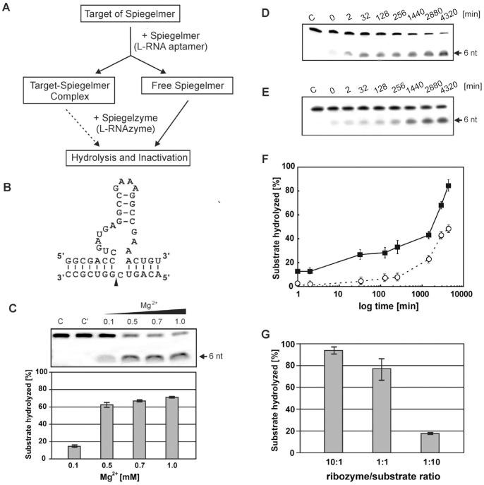
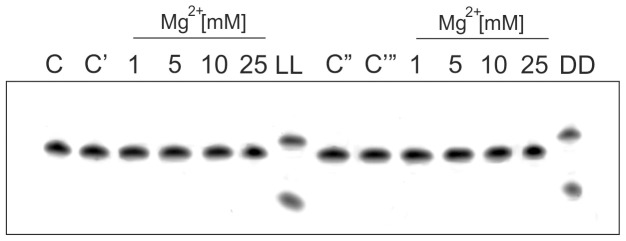
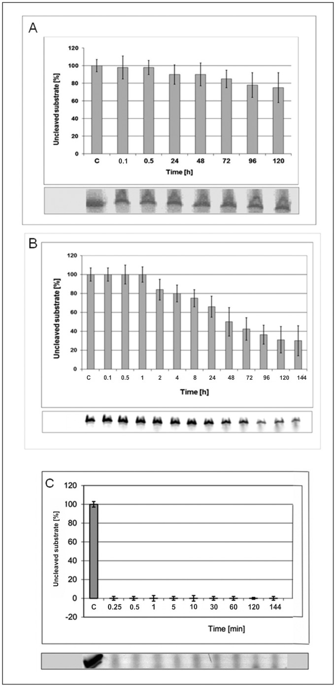
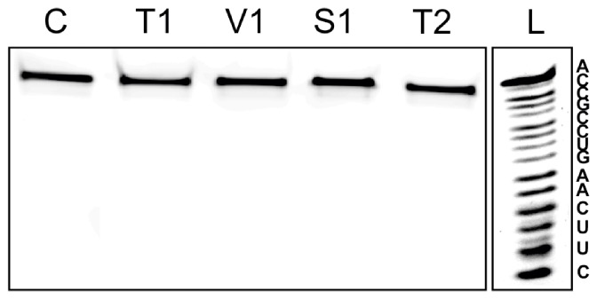
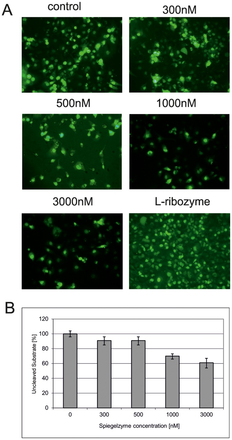
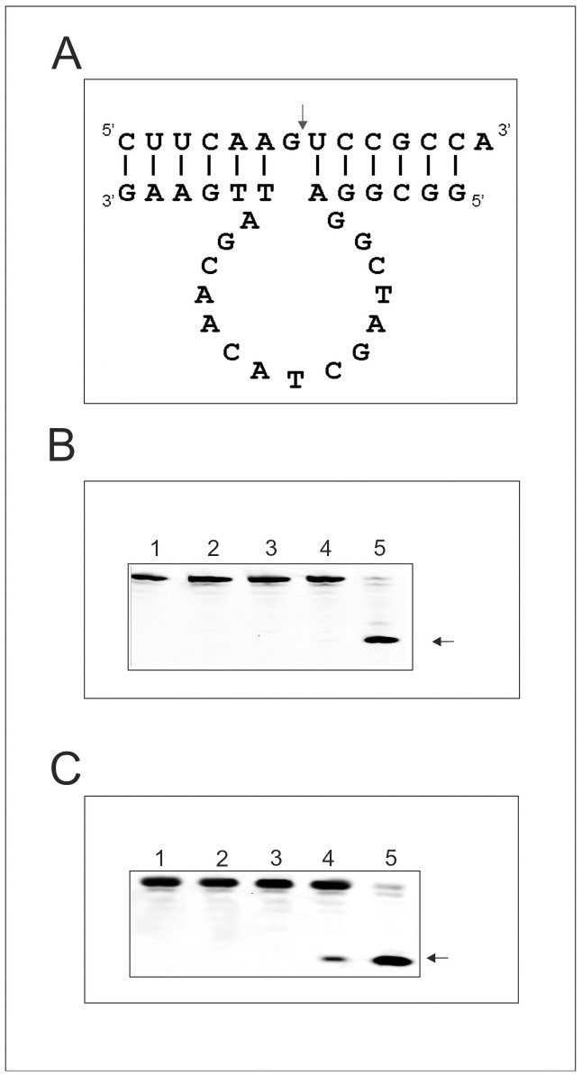
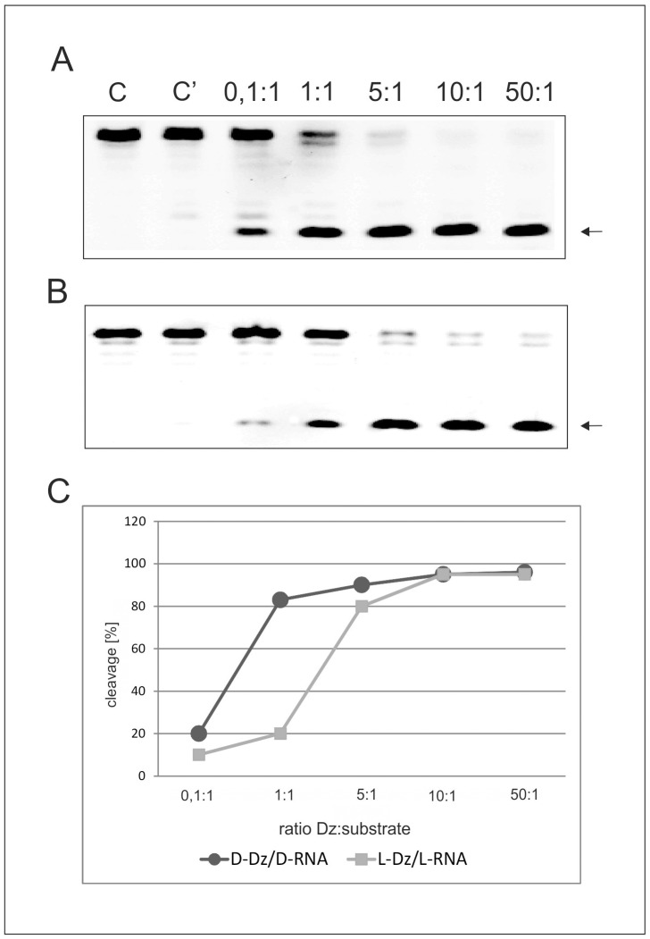
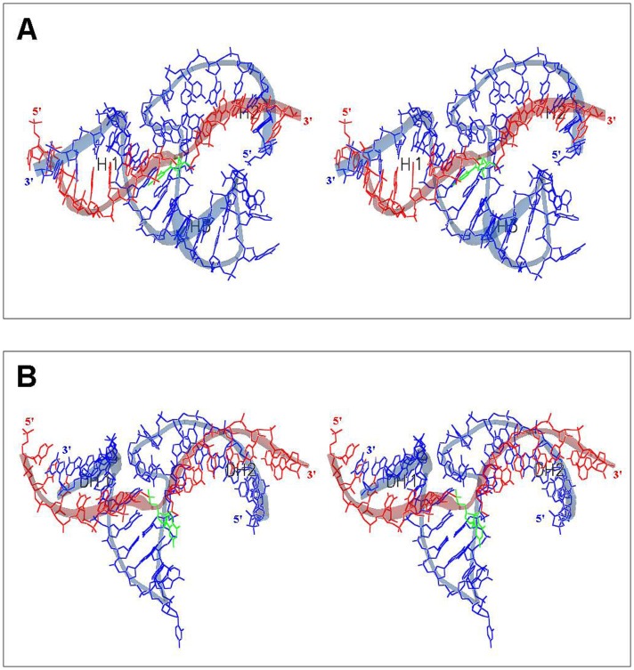
In this manuscript we describe for the first time mirror image catalytic nucleic acids (Spiegelzymes), which hydrolyze sequence specifically L-ribonucleic acid molecules. The mirror image nucleic acid ribozymes designed are based upon the known hammerhead ribozyme and DNAzyme structures that contain L-ribose or L-deoxyribose instead of the naturally occurring D-ribose or D-deoxyribose, respectively. Both Spiegelzymes show similar hydrolytic activities with the same L-RNA target molecules and they also exhibit extra ordinary stabilities when tested with three different human sera. In this respect they are very similar to Spiegelmers (mirror image aptamers), which we had previously developed and for which it has been shown that they are non-toxic and non-immunogenic. Since we are also able to demonstrate that the hammerhead and DNAzyme Spiegelzymes can also hydrolyze mirror image oligonucleotide sequences, like they occur in Spiegelmers, in vivo, it seems reasonable to assume that Spiegelzymes may in principle be used as an antidote against Spiegelmers. Since the Spiegelzymes contain the same building blocks as the Spiegelmers, it can be expected that they will have similar favorable biological characteristics concerning toxicity and immunogenety. In trying to understand the mechanism of action of the Spiegelzymes described in this study, we have initiated for the first time a model building system with L-nucleic acids. The models for L-hammerhead ribozyme and L-DNAzyme interaction with the same L-RNA target will be presented.


Conflict of interest statement

Competing Interests: The authors have the following patent applications: “Pharmazeutische Zusammensetzung zur Behandlung von Nebenwirkungen durch Gabe von Spiegelmeren” PCT/DE 2010/00159; “Pharmazeutische Zusammensetzung enthaltend L-DNA” DE 10 2010 056 610.1; and “Spiegelselektion und Spiegelevolution von Nukleinsäuren” PCT/EP 97/04726. One author (FM) is the owner of the Pentafolinum-Soft company and he is collaborating with all coauthors on the 3D modeling of Spiegelzymes. There are no further patents, products in development or marketed products to declare. This does not alter the authors’ adherence to all the PLOS ONE policies on sharing data and materials.

Figures

Figure 1
Figure 1. L-RNA1 Hydrolysis with an L-Hammerhead Ribozyme.
(A) Scheme for the potential inactivation of Spiegelmers (L-RNAs) by Spiegelzymes (L-hammerhead ribozyme). (B) The L-RNA1 and the L-hammerhead ribozyme are presented as the nucleotide sequence specific homochiral complex. The cleavage site (GUC) in the target L-RNA1 is indicated by an arrow. (C) Analysis of Spiegelmer hydrolysis at 0.1, 0.5, 0.7 and 1 mM MgCl2. Control reactions were carried out with target L-RNA1 alone in buffer and 1 mM MgCl2 (lane C) or with target L-RNA1 and L-hammerhead ribozyme in buffer without Mg2+ (lane C’). (D, E) Time dependent cleavage of the L-RNA1 target with the L-hammerhead ribozyme (Spiegelzyme). The hydrolysis of 0.2 µM target fluorescein labeled L-RNA1, with 2 µM or 0.02 µM Spiegelzyme for single (D) and multiple (E) turnover reactions. The reactions were carried out in 50 mM Tris-HCl, pH 7.5, buffer containing 1 mM MgCl2 at 37°C in total volume of 200 µl. Lanes 1–8 show the hydrolysis product (6 nt long) after 0, 2, 32, 128 and 256 min, and 24, 48 and 72 h, respectively. (F) A plot of single (squares, solid line) and multiple (open circles, broken line) turnover reactions. (G) Hydrolysis of the L-RNA1 target with the hammerhead Spiegelzyme in human serum (Sigma 2009) at different Spiegelzyme to substrate ratios (10∶1, 1∶1, 1∶10). The reactions were carried out at 37°C for 2 h. All reaction products were separated by 20% polyacrylamide gel electrophoresis (PAGE) with 7 M urea (other details see Materials and Methods) and evaluated by fluorescence measurement with Fuju Film FLA 5100 phosphoimager (F).
Figure 2
Figure 2. Hydrolysis of L-DNA1 by an L-Hammerhead Ribozyme and D-DNA1 by a D-Hammerhead Ribozyme at various Mg++ Concentrations.
Left panel: Target L-DNA1 in buffer without MgCl2 (C), the same with 25 mM of MgCl2 (C’), Target L-DNA1 with a hammerhead Spiegelzyme at 1, 5, 10 and 25 mM MgCl2, LL control: L-RNA1 incubated with L-hammerhead. Right panel: Target D-DNA1 in buffer without (C’’) and with 25 mM MgCl2 (C’’’). Target D-DNA1 and hammerhead ribozyme at 1, 5, 10 and 25 mM MgCl2, DD control: D- RNA1 incubated with D-hammerhead ribozyme. Arrow identifies hydrolysis site as in Fig. 1B.
Figure 3
Figure 3. Hammerhead Spiegelzyme (34-mer) Stability in Human Blood Sera.
(A) Incubation at 37°C for 0.25–120 h (6 days) in a human blood serum purchased from Sigma . (B) Incubation at 37°C for 0.25–144 h in serum derived from a whole blood of a healthy blood donor. Blood was collected without anticoagulant and clotting was allowed over night at room temperature. Serum was prepared by centrifugation for 30 min at 3000 rpm in a Heraeus Megafuge 3 or at 4°C und stored in aliquots at −25°C. Serum was tested negative for HIV and hepatitis B and C by routine serological tests. (C) Control-Hammerhead ribozyme (same sequence as the Spiegelzyme) was incubated in serum (as in A) at 37°C for 0.25–120 min. All diagrams show fractions of the intact L-RNA, or D-RNA at a given time point. C-control sample was incubated on ice over time period given.
Figure 4
Figure 4. Hydrolysis Experiments of L-RNA2 with T1, V1, S1, and T2 Ribonucleases.
Incubation conditions as described under Materials and Methods. L: alkaline L-RNA2 ladder. The nucleotide sequence of L-RNA2 is indicated. All nucleases were active against D-RNA (data not shown).
Figure 5
Figure 5. Hammerhead Spiegelzyme Activities in COS-7 cells.
(A) Cells were transfected with 5′-fluorescein labeled L-RNA-1 substrates. Prior to the application of the HH Spiegelzyme, the cells were washed to remove the untransfected substrate from the medium. Then the cells were transfected with 300, 500, 1000 and 3000 nM of the Spiegelzyme. The control transfection was done only with the substrate. To check for the ability of the Spiegelzyme to cross the membrane, the cells were transfected with the fluorescein labeled Spiegelzyme (bottom row, right panel). The microscopic images were taken prior to RNA isolation after washing with PBS buffer. (B) The percentage of uncleaved substrate in the L-RNA-1 isolated from cells transfected with the Spiegelzyme. After incubation for 24 hours, L-RNA was isolated from harvested cells using TRIZOL (Ambion) according to the manufacturer’s protocol. L-RNA-1 was separated by 20% PAGE with 8 M urea and the amount of substrate was determined by the fluorescence intensity using Fuji Film FLA 5100 phosphoimager.
Figure 6
Figure 6. D- or L-RNA2 Hydrolysis by D- or L-DNAzyme.
The general secondary structural model for the D- or L-DNAzyme 10/23 (A), in complex with a D- or L-RNA2 substrate (B). Hydrolysis of D-RNA2 with D-DNAzyme at 10 mM MgCl2. Lanes 1: D-RNA2 incubation in water, lane 2: D-RNA2 incubation in 50 mM Tris-HCl pH 7.5 buffer, lane 3: D-RNA2 incubation in the buffer containing in addition 10 mM MgCl2; lane 4: D-RNA2 hydrolysis with D-DNAzyme in buffer in the absence of MgCl2; lane 5: D-RNA2 hydrolysis with D-DNAzyme in buffer in the presence of 10 mM MgCl2 (C). L-RNA2 hydrolysis with L-DNAzyme at 10 mM MgCl2, analyzed with 20% PAGE with 7 M urea. Lanes 1: L-RNA2 incubation in water, lane 2: L-RNA2 incubation in 50 mM Tris-HCl pH 7.5 buffer, lane 3: L-RNA2 incubation in buffer containing in addition 10 mM MgCl2, lane 4: hydrolysis of L-RNA with L-DNAzyme in the absence of MgCl2; lane 5: hydrolysis of L-RNA substrate with L-DNAzyme in presence of 10 mM MgCl2, All incubations were carried out for 3 hrs and analyzed with 20% PAGE with 7 M urea. Arrows show the specific cleavage site in the D- and L- RNA2 targets.
Figure 7
Figure 7. Hydrolysis of D- or L-RNA2 at different D- or L- DNAzyme ratios.
Panel (A) D-DNAzyme hydrolysis of D-RNA2 at enzyme to substrate ratios as indicated in the lanes of the figure. Lane C: D-RNA2 incubated only in buffer, lane C’: D-RNA2 incubated in buffer with 10 mM MgCl2. Panel (B) L-DNAzyme hydrolysis of L-RNA2 at enzyme to substrate ratios as indicated in the lanes of the figure. Lane C: L-RNA2 incubated only in buffer, lane C’: L-RNA2 incubated in buffer with 10 mM MgCl2. (C) D-DNAzyme hydrolysis of D-RNA2 (black circles) at enzyme to substrate ratios as indicated in the figure and L-DNAzyme hydrolysis of L-RNA2 (gray squares) at ribozyme to substrate ratios shown in the panel. Incubation periods 3 h for all other conditions see Materials and Methods.
Figure 8
Figure 8. L-DNAzyme (27-mer) Stability in Different Human Sera.
Panel A: L-DNAzyme (Spiegelzyme), Panel B. D-DNAzyme and Panel C: The % fraction of D-DNAzyme degraded at given times. Sera used in these experiments Sigma 2009 (I), Sigma 1996 (II) and serum isolated from a patient (III). The DNAzymes were incubated with the sera at 37°C for the times indicated in hrs in the panels. Other details see Materials and Methods.
Figure 9
Figure 9. Atomic Models Proposed for the L-Hammerhead Ribozyme and the L-DNAzyme Interactions with their Target L-RNA2.
(A) L-HHRz, 5′-U1GGCGCUGAUGAGGCCGAAAGGCCGAAACUUGA33-3' (shown in blue) with L-RNA2 target nucleotide sequence 5′-C1UUCAAGUCCGCCA14-3′ (shown in red) with the cleavage site at nucleotide C9 (shown in green), (B) L-DNAzyme 5′-G1GCGGAGGCTAGCTACAACGATTGAAG27-3′ (shown in blue) with L-RNA2 target nucleotide sequence 5′-C1UUCAAGUCCGCCA14-3′ (shown in red) with the cleavage site at nucleotide G7 (shown in green). See text for detail discussions of the models.

References

    1. Carothers JM, Szostak JW (2006) In vitro selection of functional oligonucleotides and the origins of biochemical activity. In: The Aptamer Handbook, Functional Oligonuleotides and Their Applications, Ed. S. Klussmann, Wiley-VCH Weinheim.
    1. Xu W, Ellington AD (1996) Anti-peptide aptamers recognize amino sequence and bind a protein epitope. Proc Natl Acad Sci USA 93: 7475–7480. - PMC - PubMed
    1. Klussmann S, Nolte A, Bald R, Erdmann VA, Fürste JP (1996) Mirror-image RNA that binds D-adenosine. Nat Biotechnol 14: 1112–1115. - PubMed
    1. Nolte A, Klussmann S, Bald R, Erdmann VA, Fürste JP (1996) Mirror-design of L-oligonucleotide ligands binding to L-arginine. Nat Biotechnol 14: 1116–1119. - PubMed
    1. van Rooij E, Olson EN (2012) MicroRNA therapeutics for cardiovascular disease: opportunities and obstacles. Nat Rev Drug Discov 11: 860–872. - PMC - PubMed

Publication types