. 2013;8(1):e54914.
doi: 10.1371/journal.pone.0054914. Epub 2013 Jan 31.

Cyclophilin D deficiency rescues axonal mitochondrial transport in Alzheimer's neurons

Affiliations

Cyclophilin D deficiency rescues axonal mitochondrial transport in Alzheimer's neurons

Lan Guo et al. PLoS One. 2013.

Abstract

Normal axonal mitochondrial transport and function is essential for the maintenance of synaptic function. Abnormal mitochondrial motility and mitochondrial dysfunction within axons are critical for amyloid β (Aβ)-induced synaptic stress and the loss of synapses relevant to the pathogenesis of Alzheimer's disease (AD). However, the mechanisms controlling axonal mitochondrial function and transport alterations in AD remain elusive. Here, we report an unexplored role of cyclophilin D (CypD)-dependent mitochondrial permeability transition pore (mPTP) in Aβ-impaired axonal mitochondrial trafficking. Depletion of CypD significantly protects axonal mitochondrial motility and dynamics from Aβ toxicity as shown by increased axonal mitochondrial density and distribution and improved bidirectional transport of axonal mitochondria. Notably, blockade of mPTP by genetic deletion of CypD suppresses Aβ-mediated activation of the p38 mitogen-activated protein kinase signaling pathway, reverses axonal mitochondrial abnormalities, improves synaptic function, and attenuates loss of synapse, suggesting a role of CypD-dependent signaling in Aβ-induced alterations in axonal mitochondrial trafficking. The potential mechanisms of the protective effects of lacking CypD on Aβ-induced abnormal mitochondrial transport in axon are increased axonal calcium buffer capability, diminished reactive oxygen species (ROS), and suppressing downstream signal transduction P38 activation. These findings provide new insights into CypD-dependent mitochondrial mPTP and signaling on mitochondrial trafficking in axons and synaptic degeneration in an environment enriched for Aβ.


Conflict of interest statement

Competing Interests: The authors have declared that no competing interests exist.

Figures

Figure 1
Figure 1. Loss of CypD protects axonal mitochondrial motility and dynamics from Aβ toxicity.
(A) CypD depletion increased axonal mitochondrial density (numbers per micron of axon) in Aβ-treated neurons. rAβ: reversed Aβ42-1. There is no significant difference in the axonal mitochondrial density between vehicle-treated nonTg and Ppif −/− neurons. Data were collected from 3 independent experiments. (B) CypD depletion decreased the percentage of stationary mitochondria in Aβ-treated neurons. There were no significant changes in the percentage of stationary mitochondria between vehicle-treated nonTg and Ppif −/− neurons. Data were collected from 1380, 1074, 1410 mitochondria from vehicle, Aβ and rAβ groups in nonTg neurons, and 1634, 1505, 642 mitochondria in Ppif −/−neurons, respectively, in 4 independent experiments. (C) CypD depletion restored the decrease in the percentage of anterograde mitochondria (C1) and retrograde mitochondria (C2) in Aβ-treated neurons. Data were collected from 4 independent experiments. (D) CypD depletion increased the velocity of mitochondrial movement. (D1) Aβ treatment deceased the velocity of anterograde movement of nonTg mitochondria but not in CypD-deficient (Ppif −/−) mitochondria. Data were collected from 209, 141, 46 mitochondria from vehicle, Aβ and rAβ groups in nonTg neurons, and 158, 209, 52 mitochondria in Ppif −/−neurons. (D2–3) The cumulative distribution data showed a left shift of the velocity of anterograde mitochondria when comparing the curve for Aβ-treated nonTg mitochondrial to Ppif −/− mitochondria. Data were collected from 3 independent experiments, respectively. (D4) Aβ treatment had no effect on the velocity of the retrograde mitochondria from both nonTg and Ppif −/− mice. (E) CypD depletion rescued axonal mitochondrial mobility. Images in the top portion of the panel and kymographs in the lower panel were generated from the live imaging movies. In the kymographs, the X axis represents the mitochondrial position and the Y axis is time. Vertical white lines represent stationary mitochondria and diagonal lines represent moving mitochondria. Anterograde movements are from left to right, retrograde movements are reversed. Scale bars represent 10 µm.
Figure 2
Figure 2. Effect of CypD on Aβ-induced changes in axonal mitochondrial morphology.
(A) The average length of axonal mitochondria decreased in Aβ-treated nonTg neurons, but was largely preserved in Ppif −/− neurons. Data were collected from 3 independent experiments. (B, C) Cumulative distribution data showed that Aβ treatment caused a remarkable increase in fragmentation of small mitochondria and a decrease in the numbers of long mitochondria in nonTg neurons; this was partially attenuated in Ppif −/−neurons.
Figure 3
Figure 3. Effect of CypD depletion on Aβ-induced intra-axonal calcium elevation.
(A) Aβ-treated nonTg hippocampal neurons displayed an increase in axonal calcium levels. CypD-deficient or CsA-treated (500 nM for 24 hours) neurons diminished elevated levels of calcium. rAβ had no effect on axonal calcium levels. Data were derived from 3 independent experiments. (B) Representative images of axonal calcium staining in nonTg and Ppif −/−hippocampal neurons at indicated treatment. Scale bar represents 2 µm. (C–G2) Effect of CypD depletion on calcium ionophore (A23187)-impaired axonal mitochondrial motility. NonTg and Ppif −/−hippocampal neurons were exposed to A23187 (5 µM for 30 min) and subjected to recording of axonal mitochondrial movements including movable (C), stationary (D), anterograde (E) and retrograde (F) mitochondria. *P<0.05 vs. other groups of neurons. (G1–G2) The kymograph of axonal mitochondrial movement in nonTg (G1) and Ppif −/− (G2) neurons before and after A23187 treatment. A23187 treatment resulted in less movement than the vehicle-treated group. Ppif −/− neurons revealed increased moving traces compared to nonTg neurons in the presence of A23187. Scale bar represents 10 µm.
Figure 4
Figure 4. CypD depletion attenuates Aβ-induced intra-axonal ROS elevation.
(A) Quantification of DCF intensity in nonTg- or Ppif −/− hippocampal neurons treated with vehicle or Aβ. Addition of CsA (500 nM) to cells for 24 hours reduced the DCF intensity. Data were derived from 3 independent experiments. (B) Representative images of axonal DCF staining in nonTg and Ppif −/− hippocampal neurons for the indicated treatment. Scale bar is 10 µm. (C–D) Effect of antioxidant (Probucol) on Aβ-induced axonal mitochondrial motility. (C) Administration of Probucol (5 µM, 24 hours) ameliorated changes in Aβ-induced axonal mitochondrial motility. (D) Kymograph images show the protected effects of axonal mitochondrial moving traces following Probucol treatment. Scale bar is 10 µm.
Figure 5
Figure 5. Effect of CypD on Aβ-induced activation of p38 MAP kinase and axonal mitochondrial motility.
(A) Quantification of phospho-p38 immunoreactive bands (pT180/pY182) in hippocampal neurons treated with vehicle, Aβ, or SB203580 (SB, 1 µM) plus Aβ, respectively, which was normalized for the total p38. (B) Representative immunoblots for phospho- and total-p38. (C–E) Administration of p38 inhibitor, SB203580 (1 µM, 24 hours) to cells ameliorated Aβ-induced axonal mitochondrial motility changes (C) and mitochondrial density (D). (E) Kymographs showed the protected effects of axonal mitochondrial movement after SB203580 treatment. Scale bar is 10 µm.
Figure 6
Figure 6. Effect of CypD on Aβ-induced synaptic damage.
(A–C) Electrophysiological recording of mEPSCs for Aβ-treated nonTg and Ppif −/− neurons. CypD deficiency alleviated Aβ-induced decrease in mEPSCs frequency (A) and amplitude (B). Data were derived from 16–19 neurons for each group. (C) Representative traces of mEPSCs in the indicated group. Scale bar represents 100 pA in amplitude and 25 seconds in time. (D–E) Effect of CypD deficiency on synaptic density. The results were derived from 20–30 neurons of each group. Dendrites were visualized by the staining of MAP2 and synapses were recognized as synaptophysin-positive clusters overlapping with dendrites. (E) Representative images for double staining of synaptophysin and MAP-2 in the indicated groups. MAP2 is shown in green color and synaptophysin is labeled by red fluorescence. (F–G) Effect of Aβ-induced activation of p38 MAP kinase on synaptic density. (F) Administration of p38 inhibitor, SB203580 (1 µM, 24 hours) to cells ameliorated Aβ-induced synaptic loss. (G) Representative images showed the protected effects of synaptic density after SB203580 treatment.
Figure 7
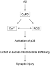
Figure 7. Working hypothesis.
Aβ-Cyclphilin D mediates impairments in axonal mitochondrial transport. In the present of Aβ, there is an increase in the opening of CypD-mediated mitochondrial permeability transition pore (mPTP), leading to disruption of Ca2+ balance and increase in reactive oxygen species (ROS) production/accumulation. Consequently, elevation of Ca2+ and oxidative activates downstream signal pathway p38 MAP Kinase contributing to mitochondrial dysfunction, deficits in axonal mitochondrial trafficking, eventually, synaptic damage.

References

    1. Kang JS, Tian JH, Pan PY, Zald P, Li C, et al. (2008) Docking of axonal mitochondria by syntaphilin controls their mobility and affects short-term facilitation. Cell 132: 137–148. - PMC - PubMed
    1. Miller KE, Sheetz MP (2004) Axonal mitochondrial transport and potential are correlated. Journal of cell science 117: 2791–2804. - PubMed
    1. Billups B, Forsythe ID (2002) Presynaptic mitochondrial calcium sequestration influences transmission at mammalian central synapses. The Journal of neuroscience : the official journal of the Society for Neuroscience 22: 5840–5847. - PMC - PubMed
    1. David G, Barrett EF (2000) Stimulation-evoked increases in cytosolic [Ca(2+)] in mouse motor nerve terminals are limited by mitochondrial uptake and are temperature-dependent. The Journal of neuroscience : the official journal of the Society for Neuroscience 20: 7290–7296. - PMC - PubMed
    1. Talbot JD, David G, Barrett EF (2003) Inhibition of mitochondrial Ca2+ uptake affects phasic release from motor terminals differently depending on external [Ca2+]. Journal of neurophysiology 90: 491–502. - PubMed

Publication types

MeSH terms

Substances