. 2013;8(1):e54960.
doi: 10.1371/journal.pone.0054960. Epub 2013 Jan 28.

Pencilbeam irradiation technique for whole brain radiotherapy: technical and biological challenges in a small animal model

Affiliations

Pencilbeam irradiation technique for whole brain radiotherapy: technical and biological challenges in a small animal model

Elisabeth Schültke et al. PLoS One. 2013.

Abstract

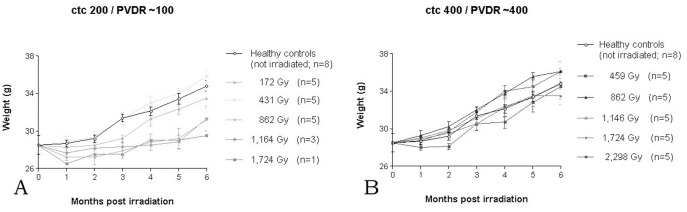
We have conducted the first in-vivo experiments in pencilbeam irradiation, a new synchrotron radiation technique based on the principle of microbeam irradiation, a concept of spatially fractionated high-dose irradiation. In an animal model of adult C57 BL/6J mice we have determined technical and physiological limitations with the present technical setup of the technique. Fifty-eight animals were distributed in eleven experimental groups, ten groups receiving whole brain radiotherapy with arrays of 50 µm wide beams. We have tested peak doses ranging between 172 Gy and 2,298 Gy at 3 mm depth. Animals in five groups received whole brain radiotherapy with a center-to-center (ctc) distance of 200 µm and a peak-to-valley ratio (PVDR) of ∼ 100, in the other five groups the ctc was 400 µm (PVDR ∼ 400). Motor and memory abilities were assessed during a six months observation period following irradiation. The lower dose limit, determined by the technical equipment, was at 172 Gy. The LD50 was about 1,164 Gy for a ctc of 200 µm and higher than 2,298 Gy for a ctc of 400 µm. Age-dependent loss in motor and memory performance was seen in all groups. Better overall performance (close to that of healthy controls) was seen in the groups irradiated with a ctc of 400 µm.


Conflict of interest statement

Competing Interests: The authors have declared that no competing interests exist.

Figures

Figure 1
Figure 1. Schematic comparison between the profile of energy deposition in the direction of beam propagation with microplanar beams as in the classic MRT approach (A) and in pencilbeam irradiation (B).
Based on the geometry of beam deposition we would expect a higher tolerance of high dose irradiation in the normal tissue for the pencilbeam approach (less interruption of normal structure).
Figure 2
Figure 2. Experimental setup for pencilbeam irradiation.
The animal was positioned orthogonally to the direction of beam propagation (right to left) and dose delivery was verified using Gafchromic film, visible in right lateral position. For pencilbeam irradiation, the goniometer with the animal was moved vertically through the beam, along the axis marked by the broken arrow.
Figure 3
Figure 3. Beam profiles for ctc 200 µm (A) and ctc 400 µm (B) at 3 mm depth.
Profiles parallel and perpendicular to the polarisation vector are shown. The FWHM is 55 µm.
Figure 4
Figure 4. Peak and Valley doses in dependency on depth in the centre of the pencil beam field.
The irradiation field contains 45×21 pencil beams and the phantom is approximated as a homogeneous box of water with 8 cm side length. Valley dose can be measured vertically or laterally between two pencil beams and diagonal in the centre between two beams. The latter gives the lowest value due to the greatest distance from the beams. The other two valley doses differ slightly due to polarisation effects.
Figure 5
Figure 5. Weight curves for the animals in the two beam configurations tested, ctc 200 µm (A) and ctc 400 µm (B).
The irradiation doses in the legends are given as peak dose.
Figure 6
Figure 6. Duration of performance on the rotarod over the 6 months observation period.
Performance was generally better in the ctc 400 µm groups with a PVDR of ∼ 400 (B), compared to the animals in the ctc 200 µm groups with a PVDR of ∼ 100 (A).
Figure 7
Figure 7. Ability to form new memory as assessed by Object Recognition Test (ORT).
Performance was generally better in the ctc 400 µm groups (B), compared to the animals in the ctc 200 µm groups (A). The initial drop of memory performance is lagging behind that of motor performance by several weeks.
Figure 8
Figure 8. H&E histology, sagittal sections of the cerebellum 6 months after irradiation.
C57 BL/J6 mice, 6 months after irradiation with a peak dose of 172 Gy/valley dose of 1.72 Gy, ctc 200 µm (A, C-F are enlargements from sample A) and healthy control (B). The lighter spots almost devoid of cells correspond to the ctc 200 µm lateral Gafchromic film profile as can be demonstrated by the overlaid patterns of histology section and distance grid with 200 µm lateral dimensions (D and F). Because the geometry of mounted tissue sections is influenced by several aspects of the preparation process, the histological pattern does not perfectly correspond to the pattern recorded on Gafchromic film (i.e. the grid lines are not quite orthogonal).
Figure 9
Figure 9. H&E histology of C57 BL/J6 mice, axial sections 6 months after irradiation.
Showing a 5 µm thick axial section after irradiation with a peak dose of 1,164 Gy and a valley dose 11.64 Gy, ctc 200 µm (A) and the axial histology section overlaid with the photo of the lateral beam profile (B). Only a few cells, some of them with small dark (pyknotic) nuclei, are left in the path of the beam.

Similar articles

Cited by

References

    1. Bräuer-Krisch E, Requardt H, Brochard T, Berruyer G, Renier M, et al. (2009) New technology enables high precision multislit collimators for microbeam radiation therapy, Rev Sci Instr. 80: 074301. - PubMed
    1. Laissue JA, Geiser G, Spanne PO, Dilmanian FA, Gebbers JO (1998) Neuropoathology of ablation of rat gliosarcomas and contiguous brain tissues using a microplanar beam of synchrotron-wiggler-generated x-rays. Int J Cancer 78: 654–660. - PubMed
    1. Schültke E, Juurlink B, Ataelmannan K, Laissue J, Blattmann H, et al. (2008) Memory and Survival after Microbeam Radiation Therapy. Eur J Radiol 68S: S142–S146. - PubMed
    1. Laissue JA, Blattmann H, Di Michiel M, Slatkin DN, Lyubimov N, et al. (2001) Weanling piglet cerebellum: a surrogate for tolerance to MRT (microbeam radiation therapy) in pediatric neuro-oncology. Proc SPIE 4508: 65–73.
    1. Bouchet A, Lemasson B, Le Duc G, Maisin C, Bräuer-Krisch E, et al. (2010) Preferential effect of synchrotron microbeam radiation therapy on intracerebral 9L gliosarcoma vascular networks. Int J Radiat Oncol Biol Phys 78(5): 1503–12. - PubMed

Publication types