. 2013;8(1):e55018.
doi: 10.1371/journal.pone.0055018. Epub 2013 Jan 28.

Dengue virus enhances thrombomodulin and ICAM-1 expression through the macrophage migration inhibitory factor induction of the MAPK and PI3K signaling pathways

Affiliations

Dengue virus enhances thrombomodulin and ICAM-1 expression through the macrophage migration inhibitory factor induction of the MAPK and PI3K signaling pathways

Trai-Ming Yeh et al. PLoS One. 2013.

Abstract

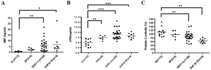
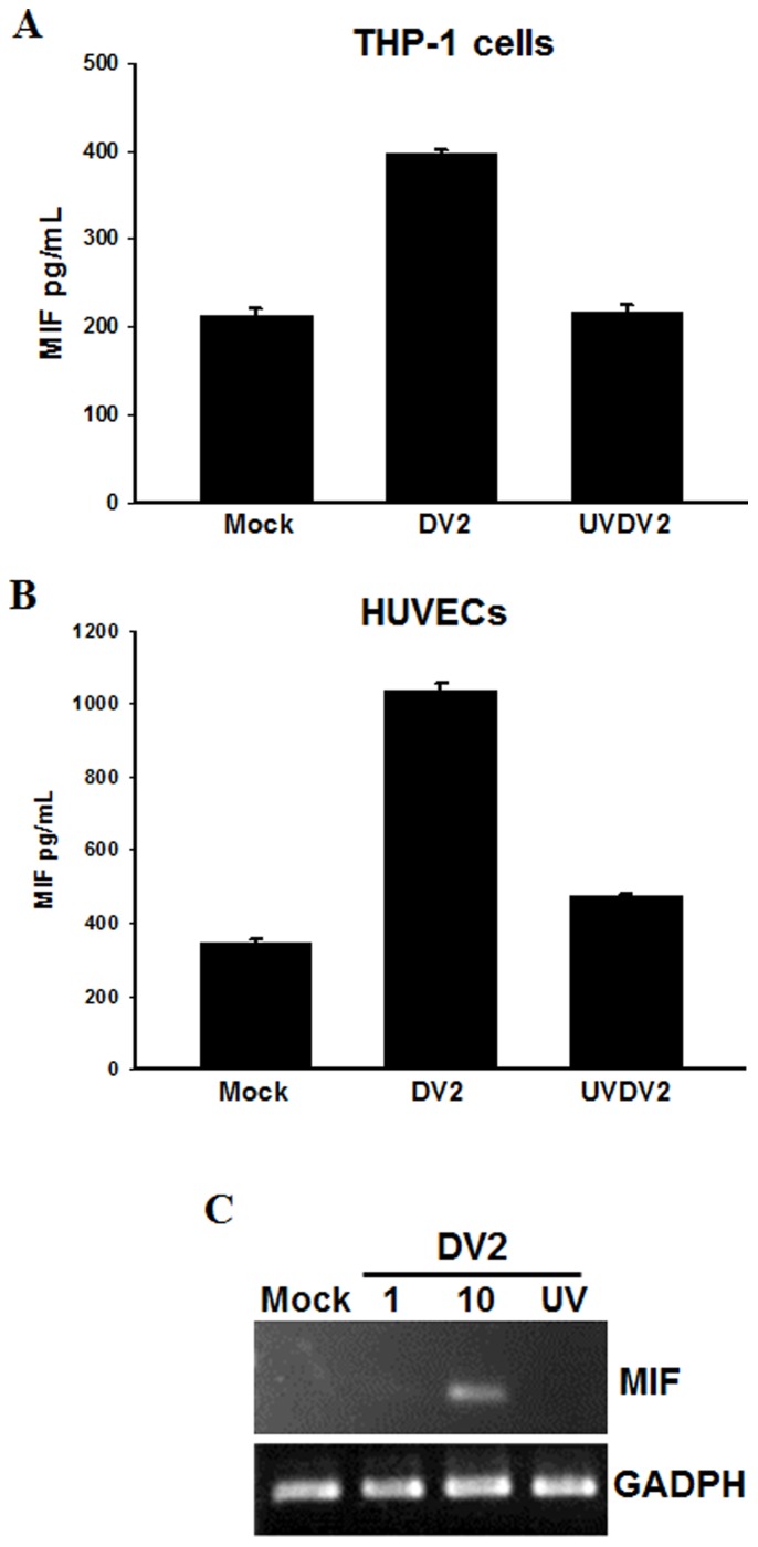
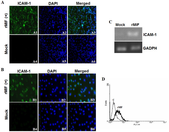
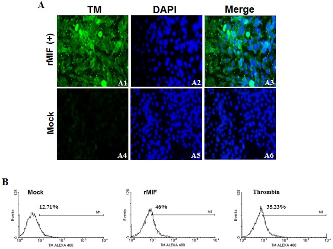
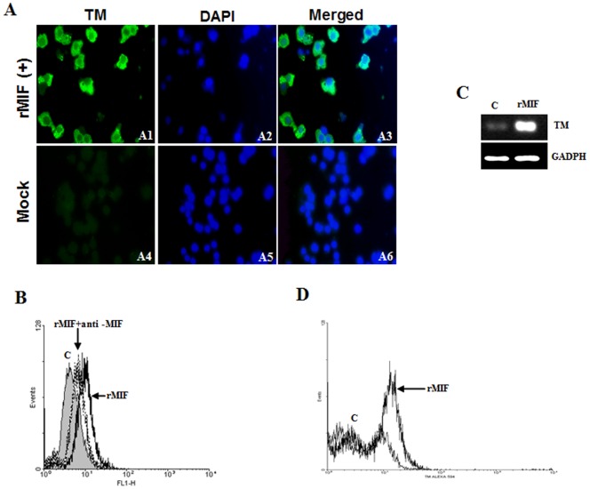
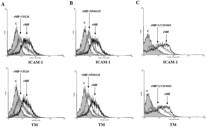
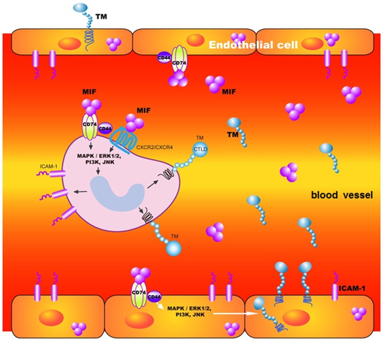
Dengue virus (DV) infections cause mild dengue fever (DF) or severe life-threatening dengue hemorrhagic fever (DHF). The mechanisms that cause hemorrhage in DV infections remain poorly understood. Thrombomodulin (TM) is a glycoprotein expressed on the surface of vascular endothelial cells that plays an important role in the thrombin-mediated activation of protein C. Prior studies have shown that the serum levels of soluble TM (sTM) and macrophage migration inhibitory factor (MIF) are significantly increased in DHF patients compared to levels in DF patients or normal controls. In this study, we investigated how MIF and sTM concentrations are enhanced in the plasma of DHF patients and the potential effect of MIF on coagulation through its influence on two factors: thrombomodulin (TM) and intercellular adhesion molecule-1 (ICAM-1) in endothelial cells and monocytes. Recombinant human macrophage migration inhibitory factor (rMIF) was used to treat monocytic THP-1 cells and endothelial HMEC-1 cells or primary HUVEC cells. The subsequent expression of TM and ICAM-1 was assessed by immunofluorescent staining and flow cytometry analysis. Additionally, the co-incubation of THP-1 cells with various cell signaling pathway inhibitors was used to determine the pathways through which MIF mediated its effect. The data provided evidence that severe DV infections induce MIF expression, which in turn stimulates monocytes or endothelial cells to express TM and ICAM-1 via the Erk, JNK MAPK and the PI3K signaling pathways, supporting the idea that MIF may play an important role as a regulator of coagulation.


Conflict of interest statement

Competing Interests: The authors have declared that no competing interests exist.

Figures

Figure 1
Figure 1. MIF, sTM and PC concentrations are increased in plasma of patients with DHF.
Levels of (A) MIF, (B) soluble thrombomodulin (sTM) and (C) Protein C (PC) in patients infected with dengue virus and in control groups. Sera from healthy controls (N, n = 12), dengue fever (DF) patients (n = 12) and dengue hemorrhagic fever patients (DHF) sorted into grades I–II (n = 28) and grades III–IV (n = 9) were assayed for MIF, sTM and PC. The amounts of MIF, sTM and PC in dengue patients were measured using ELISA kits as described in the Materials and Methods. *p<0.1, **p<0.01 and ***p<0.001 compared with corresponding values from normal controls (N). Levels of MIF and sTM were higher than in normal controls, but PC was lower than in the control.
Figure 2
Figure 2. Dengue virus induces MIF production in monocytes and human umbilical cord vein endothelial cells (HUVECs).
THP-1 cells or primary HUVECs were infected with DV2 (MOI = 10). (A, B) The concentrations of MIF in culture media were assayed at 48 h post-infection using ELISA kits as described in the Materials and Methods. Controls were uninfected cells (Mock) and cells treated with UV-inactivated DV (UVDV2). (C) DV2 induced MIF mRNA expression in HUVECs. HUVECs were infected with DV2 (MOI = 1 or 10) and incubated for 6 hours. RNA was extracted and the expression of MIF was analyzed by semi-quantitative RT-PCR with specific primers for MIF. The gene expression of GADPH was used as the internal control. Controls were cells without virus (Mock) and cells treated with UV-inactivated DV (UV).
Figure 3
Figure 3. rMIF induces ICAM-1 expression in endothelial cells and monocytes.
(A) HMEC-1 (1×10⁶) and (B) HUVEC (1×10⁶) endothelial cells were stimulated with rMIF (0.4 µg/mL) for 24 h. ICAM-1 and nuclei were stained with FITC conjugated anti-ICAM-1 antibody and DAPI, respectively, and observed using fluorescence microscopy at 400× magnification. Negative controls were treated with 0.9% saline (Mock). Thrombin-treated HMEC-1 cells were used as a positive control. THP-1 monocytic cells (2×10⁶) (C and D) were stimulated with rMIF (0.4 µg/mL) for 24 h. (C) RNA was extracted and ICAM-1 expression was analyzed using a semi-quantitative RT-PCR with specific primers for ICAM-1. The gene expression of GADPH was used as the internal control. (D) The expression of ICAM-1 was detected by flow cytometry analysis and compared to the control (without rMIF treatment). Flow cytometry analysis showed increased ICAM-1 expression in THP-1 monocytes following rMIF treatment.
Figure 4
Figure 4. rMIF induces thrombomodulin (TM) expression in endothelial cells.
(A) HMEC-1 (1×10⁶) cells were stimulated with rMIF (0.4 µg/mL) for 24 h. TM and nuclei were stained with FITC conjugated anti-TM antibody and DAPI, respectively, and observed using fluorescence microscopy at 400× magnification. The controls were treated with 0.9% saline (Mock). (B) The expression of TM in HMEC-1 cells with/without rMIF treatment was determined using flow cytometry analysis. Cells treated with rMIF showed increased TM expression compared to the control (without). The thrombin-treated cells were used as a positive control.
Figure 5
Figure 5. rMIF induces thrombomodulin (TM) expression in monocytes.
(A) THP-1 cells (2×10⁶) were stimulated with rMIF (0.4 µg/mL) for 24 h. TM and nuclei were stained with FITC conjugated anti-TM antibody and DAPI, respectively, and observed using fluorescence microscopy at 400× magnification. The controls were treated with 0.9% saline (Mock). (B) Flow cytometry analysis of TM expression in THP-1 cells with/without rMIF treatment. A neutralizing anti-MIF mAb (20 µg/mL) was used to abrogate the MIF-induced TM expression. (C) RNA was extracted and TM expression was analyzed through a semi-quantitative RT-PCR with specific primers for TM. The GADPH was used as the internal control. TM expression was enhanced with rMIF treatment in THP-1 cells, compared to the control (c; without rMIF treatment). (D) Flow cytometry analysis also showed increased TM expression in PBMCs following rMIF treatment.
Figure 6
Figure 6. rMIF enhances ICAM-1 and TM expression via the Erk, JNK MAPK and the PI3K signaling pathways in THP-1 cells.
(A) Erk inhibitor U0126, (B) JNK inhibitor SP60125, or (C) PI3K inhibitor LY294002 at 20 µM in DMSO was added to the THP-1 cell culture 30 min before and throughout rMIF treatment. The cells (2×10⁶) were stimulated with rMIF (0.4 µg/mL) for 24 h. The expression of ICAM-1 (Left panel) and TM (Right panel) was assessed by flow cytometry analysis. The rMIF-treated cells were compared to the controls.
Figure 7
Figure 7. Schematic diagram of the roles of MIF in DV-infected patients’ circulation.
DV can stimulate monocytes and endothelial cells to express MIF. MIF signals through binding to a functional receptor complex CD74/CD44 and G-protein-coupled chemokine receptors (CXCR2/CXCR4) individually . Following signaling may activate PI3K/MEK/ERK and downstream JNK signaling pathways, resulting in TM and ICAM-1 expression and causing soluble TM levels to increase in plasma.

References

    1. Gubler DJ (1998) Dengue and dengue hemorrhagic fever. Clin Microbiol Rev 11: 480–496. - PMC - PubMed
    1. Mairuhu AT, Mac Gillavry MR, Setiati TE, Soemantri A, ten Cate H, et al. (2003) Is clinical outcome of dengue-virus infections influenced by coagulation and fibrinolysis? A critical review of the evidence. Lancet Infect Dis 3: 33–41. - PubMed
    1. Huang YH, Liu CC, Wang ST, Lei HY, Liu HL, et al. (2001) Activation of coagulation and fibrinolysis during dengue virus infection. J Med Virol 63: 247–251. - PubMed
    1. van Gorp EC, Minnema MC, Suharti C, Mairuhu AT, Brandjes DP, et al. (2001) Activation of coagulation factor XI, without detectable contact activation in dengue haemorrhagic fever. Br J Haematol 113: 94–99. - PubMed
    1. Suharti C, van Gorp EC, Setiati TE, Dolmans WM, Djokomoeljanto RJ, et al. (2002) The role of cytokines in activation of coagulation and fibrinolysis in dengue shock syndrome. Thromb Haemost 87: 42–46. - PubMed

Publication types

MeSH terms

Substances