. 2013;8(1):e55375.
doi: 10.1371/journal.pone.0055375. Epub 2013 Jan 31.

Triggering of inflammasome by aggregated α-synuclein, an inflammatory response in synucleinopathies

Affiliations

Triggering of inflammasome by aggregated α-synuclein, an inflammatory response in synucleinopathies

Gaia Codolo et al. PLoS One. 2013.

Abstract

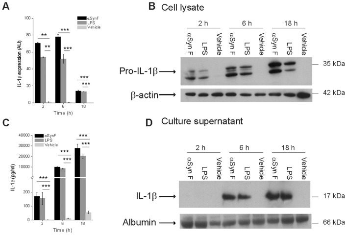
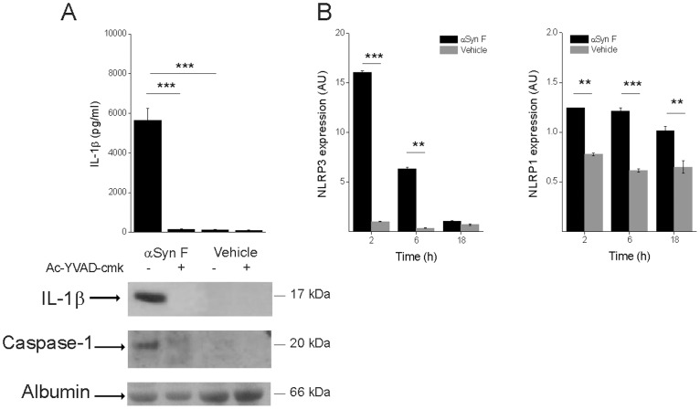
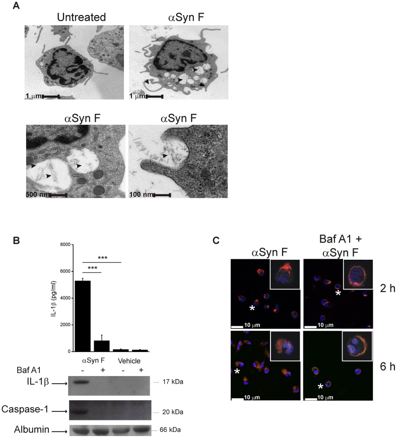
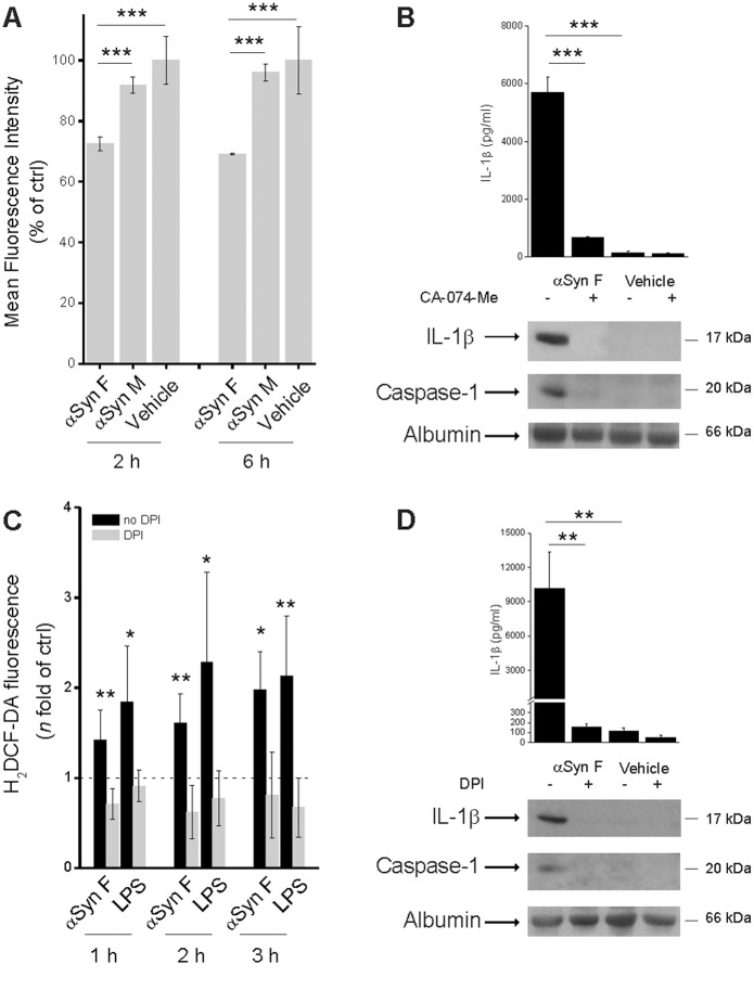
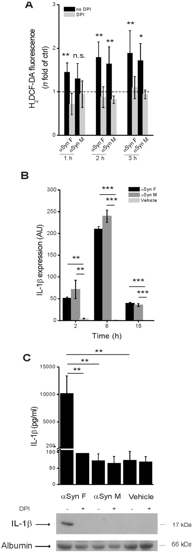
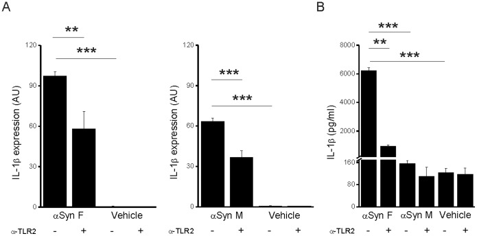
Parkinson's disease (PD) is one of the most common neurodegenerative diseases. It is characterized by the loss of dopaminergic neurons in the substantia nigra pars compacta of the brain. Another feature is represented by the formation in these cells of inclusions called Lewy bodies (LB), principally constituted by fibrillar α-synuclein (αSyn). This protein is considered a key element in the aetiology of a group of neurodegenerative disorders termed synucleinopathies, which include PD, but the cellular and molecular mechanisms involved are not completely clear. It is established that the inflammatory process plays a crucial role in the pathogenesis and/or progression of PD; moreover, it is known that aggregated αSyn, released by neurons, activates microglia cells to produce pro-inflammatory mediators, such as IL-1β. IL-1β is one of the strongest pro-inflammatory cytokines; it is produced as an inactive mediator, and its maturation and activation requires inflammasome activation. In particular, the NLRP3 inflammasome is activated by a wide variety of stimuli, among which are crystallized and particulate material. In this work, we investigated the possibility that IL-1β production, induced by fibrillar αSyn, is involved the inflammasome activation. We demonstrated the competence of monomeric and fibrillar αSyn to induce synthesis of IL-1β, through TLR2 interaction; we found that the secretion of the mature cytokine was a peculiarity of the fibrillated protein. Moreover, we observed that the secretion of IL-1β involves NLRP3 inflammasome activation. The latter relies on the phagocytosis of fibrillar αSyn, followed by increased ROS production and cathepsin B release into the cytosol. Taken together, our data support the notion that fibrillar αSyn, likely released by neuronal degeneration, acts as an endogenous trigger inducing a strong inflammatory response in PD.


Conflict of interest statement

Competing Interests: The authors have declared that no competing interests exist.

Figures

Figure 1
Figure 1. IL-1β synthesis and secretion in human monocytes induced by fibrillar αSyn.
(A) Monocytes were exposed for 2, 6 and 18 h to 40 nM αSyn F or 1 µg/ml LPS (positive control) or vehicle (saline) and the expression of pro-IL-1β was evaluated by real-time PCR. Real time data are shown as the mean ± S.D. of results obtained with cell preparations from 5 different donors; experiments with each cell preparation were conducted in duplicate. **p<0.01, ***p<0.001. (B) IL-1β synthesis was also evaluated by immunoblot analysis in cell lysates from monocytes treated as in A. The immunoblot is from one representative donor. (C) Culture supernatants from monocytes that had been harvested for quantification of mRNA levels, reported in A, were collected and evaluated for their IL-1β content by ELISA. Results are the mean ± S.D. of 5 determinations made in duplicate with different cell preparations derived from different donors. ***p<0.001 (D) Culture supernatants of monocytes, harvested for the evaluation of IL-1β synthesis, as in B, were collected and the released IL-1β revealed in immunoblot.
Figure 2
Figure 2. IL-1β induced by αSyn F is mediated by caspase-1 activation and involves NLRP3.
(A) Monocytes were pre-incubated for 30 min with 50 µM Ac-YVAD-cmk, or left untreated, before being exposed for 6 h to 40 nM αSyn F or vehicle. IL-1β released into the culture supernatants was evaluated both by ELISA and immunoblot analysis. The same supernatants were assessed for the accumulation of active caspase-1 by immunoblot. Results are the mean ± S.D. of 3 experiments conducted in duplicate with cell preparations obtained from 3 different donors. The immunoblot is from one representative donor. ***p<0.001 (B) Real-time PCR of NLRP1 and NLRP3 in monocytes stimulated for 2, 6 and 18 h with αSyn F. Real time data are shown as the mean ± S.D. of results obtained with cell preparations obtained from 2 different donors; experiments with each cell preparation were conducted in duplicate. **p<0.01 and ***p<0.001.
Figure 3
Figure 3. Phagocytosis of αSyn F is required for IL-1β release.
(A) Transmission electron microscopy of monocytes treated for 6 h with 40 nM αSyn F, or left untreated. For αSyn F-treated cells, three magnifications are shown (bars represent 1 µm, 500 nm and 100 nm). Pictures of 20–30 cells for each condition were kept and 3 representative cells are shown. Left bottom panel is the magnification of two αSyn-containing vacuoles of the cell in the right upper panel. Arrowheads indicate the fibrils. (B) Monocytes were pre-incubated for 30 min with 250 nM Baf A1, or left untreated, before being exposed for 6 h to αSyn F or vehicle. IL-1β released into the culture supernatants was evaluated both by ELISA and immunoblot analysis. The same supernatants were assessed for the accumulation of active caspase-1 by immunoblot. Results are the mean ± S.D. of 3 experiments conducted in duplicate with cell preparations obtained from 3 different donors. The immunoblot is from one representative donor. ***p<0.001. (C) Confocal microscopy of monocytes incubated with 250 nMBaf A1 before and during the exposure for 2 or 6 h to αSyn F. Red staining corresponds to rhodamine-labelled αSyn F, while the Hoechst blue staining labels nuclei. White asterisks indicate the cells of which a magnification is shown (white square).
Figure 4
Figure 4. Cathepsin B activity as well as ROS production are involved in αSyn F-induced inflammasome activation.
(A) Phagocytosis of fibrillar αSyn leads to phago-lysosomal destabilization. Flow cytometry of monocytes stained with acridine orange and then treated for 2 or 6 h with 40 nM αSyn F, 1 µM αSyn M. Data are reported as the percentage of acridine orange-positive cells present in the vehicle-exposed sample. Results are the mean ± S.D. of 3 experiments conducted in duplicate with cell preparations obtained from 3 different donors. ***p<0.001. (B) Monocytes were incubated for 30 min with 10 µM CA-074-Me (an inhibitor of cathepsin B), or left untreated; cells were then exposed for 6 h to 40 nM αSyn F or vehicle. IL-1β released into the culture supernatants was evaluated both by ELISA and immunoblot analysis. The same supernatants were assessed for the accumulation of active caspase-1 by immunoblot. Results are the mean ± S.D. of 3 experiments conducted in duplicate with cell preparations obtained from 3 different donors. The immunoblot is from one representative donor. ***p<0.001. (C) Monocytes were cultured for 1, 2 or 3 h in the presence of 1 µg/ml LPS or 40 nM αSyn F and intracellular ROS levels were quantified by the H2DCF-DA fluorometric method. When required, monocytes were pre-incubated for 30 min with 20 µM DPI. The dotted line refers to the basal levels of ROS production (as in untreated monocytes). Results are the mean ± S.D. of 5 experiments conducted in duplicate with cell preparations obtained from 5 different donors. *p<0.05, **p<0.01 was calculated for samples not exposed to DPI vs the correspondent samples with DPI. (D) Monocytes were pre-incubated for 30 min with 20 µM DPI, or left untreated, before being exposed for 6 h to αSyn F or vehicle. IL-1β, as well as activated caspase-1, were assessed as before. Results are the mean ± S.D. of 3 experiments conducted in duplicate with cell preparations obtained from 3 different donors. The immunoblot is from one representative donor. **p<0.01.
Figure 5
Figure 5. Only αSyn F is capable of activating the inflammasome.
(A) Monocytes were cultured for 1, 2 or 3 h in the presence of 40 nM αSyn F or 1 µM αSyn M and intracellular ROS levels were quantified by the H2DCF-DA fluorometric method. When required, monocytes were pre-incubated for 30 min with 20 µM DPI. The dotted line refers to the basal levels of ROS production (as in untreated monocytes). Results are the mean ± S.D. of 5 experiments conducted in duplicate with cell preparations obtained from 5 different donors. *p<0.05, **p<0.01 was calculated for samples not exposedto DPI vs the correspondent samples with DPI. (B) Monocytes were exposed for 2, 6 and 18 h to 40 nM αSyn F, or 1 µM αSyn M or vehicle (saline) and the expression of pro-IL-1β was evaluated by real-time PCR. Real time data are shown as the mean ± S.D. of results obtained with cell preparations from 2 different donors; experiments with each cell preparation were conducted in duplicate. **p<0.01 and ***p<0.001. (C) Monocytes were pre-incubated for 30 min with 20 µM DPI, or left untreated, before being exposed for 6 h to αSyn F or αSyn M or vehicle. IL-1β was determined as before. Results are the mean ± S.D. of 3 experiments conducted in duplicate with cell preparations obtained from 3 different donors. The immunoblot is from one representative donor. **p<0.01.
Figure 6
Figure 6. TLR2 engagement is involved in IL-1β induction by αSyn.
(A) Levels of pro-IL-1β mRNA in monocytes were determined by quantitative real-time PCR analysis after a 6 h stimulation with αSyn F or αSyn M in the presence or not of a soluble anti-TLR2 antibody. Real time data are shown as the mean ± S.D. of results obtained with 2 cell preparations obtained from 2 different donors; experiments with each cell preparation was conducted in duplicate. (B) IL-1β content in the culture supernatant of the same cells harvested for mRNA evaluation was determined by ELISA. Results are the mean ± S.D. of 2 experiments conducted in duplicate with cell preparations obtained from 2 different donors. **p<0.01 and ***p<0.001.

References

    1. de Rijk MC, Launer LJ, Berger K, Breteler MM, Dartigues JF, et al. (2000) Prevalence of Parkinson's disease in Europe: A collaborative study of population-based cohorts. Neurologic Diseases in the Elderly Research Group. Neurology 54: S21–23. - PubMed
    1. Braak H, Del Tredici K, Rub U, de Vos RA, Jansen Steur EN, et al. (2003) Staging of brain pathology related to sporadic Parkinson's disease. Neurobiol Aging 24: 197–211. - PubMed
    1. Croisier E, Moran LB, Dexter DT, Pearce RK, Graeber MB (2005) Microglial inflammation in the parkinsonian substantia nigra: relationship to alpha-synuclein deposition. J Neuroinflammation 2: 14. - PMC - PubMed
    1. Spillantini MG, Crowther RA, Jakes R, Hasegawa M, Goedert M (1998) alpha-Synuclein in filamentous inclusions of Lewy bodies from Parkinson's disease and dementia with lewy bodies. Proc Natl Acad Sci U S A 95: 6469–6473. - PMC - PubMed
    1. Gasser T (2005) Genetics of Parkinson's disease. Curr Opin Neurol 18: 363–369. - PubMed

Publication types

MeSH terms