. 2013;8(1):e55697.
doi: 10.1371/journal.pone.0055697. Epub 2013 Jan 31.

A study on the interaction of rhodamine B with methylthioadenosine phosphorylase protein sourced from an Antarctic soil metagenomic library

Affiliations

A study on the interaction of rhodamine B with methylthioadenosine phosphorylase protein sourced from an Antarctic soil metagenomic library

Paulina Bartasun et al. PLoS One. 2013.

Abstract

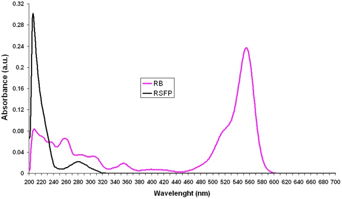
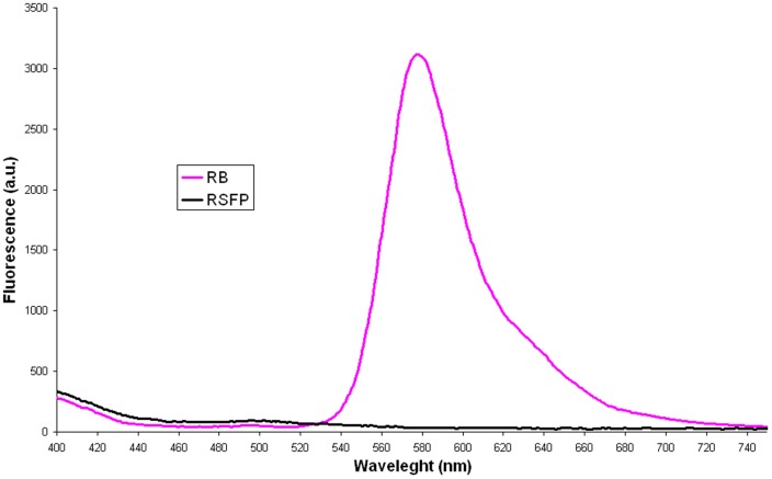
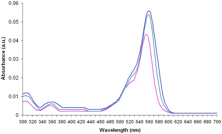
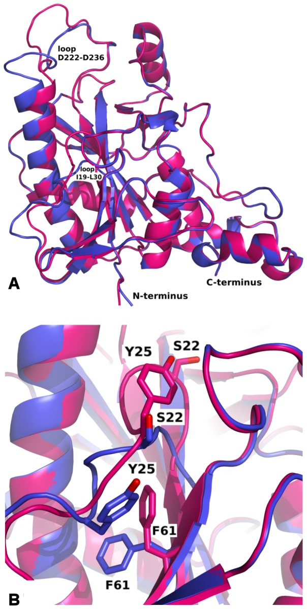
The presented study examines the phenomenon of the fluorescence under UV light excitation (312 nm) of E. coli cells expressing a novel metagenomic-derived putative methylthioadenosine phosphorylase gene, called rsfp, grown on LB agar supplemented with a fluorescent dye rhodamine B. For this purpose, an rsfp gene was cloned and expressed in an LMG194 E. coli strain using an arabinose promoter. The resulting RSFP protein was purified and its UV-VIS absorbance spectrum and emission spectrum were assayed. Simultaneously, the same spectroscopic studies were carried out for rhodamine B in the absence or presence of RSFP protein or native E. coli proteins, respectively. The results of the spectroscopic studies suggested that the fluorescence of E. coli cells expressing rsfp gene under UV illumination is due to the interaction of rhodamine B molecules with the RSFP protein. Finally, this interaction was proved by a crystallographic study and then by site-directed mutagenesis of rsfp gene sequence. The crystal structures of RSFP apo form (1.98 Å) and complex RSFP/RB (1.90 Å) show a trimer of RSFP molecules located on the crystallographic six fold screw axis. The RSFP complex with rhodamine B revealed the binding site for RB, in the pocket located on the interface between symmetry related monomers.


Conflict of interest statement

Competing Interests: The authors have declared that no competing interests exist.

Figures

Figure 1
Figure 1. SDS-PAGE analysis of the cell extract with RSFP protein and the purified enzyme.
Protein weight marker (lane M), cell extract of E. coli LMG194/pBADRSFP (lane A), pooled fraction after Fractogel EMD DEAE chromatography (lane B), pooled fraction after Resource Q chromatography (lane C).
Figure 2
Figure 2. The pink fluorescence assay: RB in PBS buffer (A), PBS buffer (B), RB+RSFP in PBS buffer (C), RSFP in PBS buffer (D), RB+E. coli LMG194/pBADRSFP cell lysate in PBS buffer (E), E. coli LMG194/pBADRSFP cell lysate in PBS buffer (F), RB+E. coli LMG194/pBADMycHisA cell lysate in PBS buffer (G), and E. coli LMG194/pBADMycHisA cell lysate in PBS buffer (H).
Figure 3
Figure 3. The UV-VIS absorption spectra of rhodamine B (RB; 1.75×10⁻³ g L−1) and RSFP protein (0.023 g L−1).
Figure 4
Figure 4. The emission spectra of rhodamine B (RB; 1.225×10⁻³ g L−1) and RSFP protein (0.081 g L−1).
Figure 5
Figure 5. The UV-VIS absorption spectra of rhodamine B (green and blue lines) in the presence of the different concentration of RSFP protein, respectively, and the UV-VIS absorption spectrum of rhodamine B in the absence of RSFP protein (pink line).
The concentration of RSFP protein in the first analyzed sample (green line) was twice higher than the concentration of RSFP protein (0.023 g L−1) in the second one (blue line), respectively. The concentration of RB (3.5×10⁻⁴ g L−1) was the same in the all assayed samples.
Figure 6
Figure 6. The UV-VIS absorption spectra of rhodamine B (brown and orange lines) in the presence of the different concentration of E. coli proteins, respectively, and the UV-VIS absorption spectrum of rhodamine B in the absence of E. coli proteins (pink line).
The concentration of E. coli proteins in the first analyzed sample (brown line) was twice higher than the concentration of E. coli proteins (0.023 g L−1) in the second one (orange line), respectively. The concentration of RB (3.5×10⁻⁴ g L−1) was the same in the all assayed samples.
Figure 7
Figure 7. The emission spectra of rhodamine B (RB) in the presence of the different concentration of RSFP protein or E. coli proteins (Lys).
The concentrations of RSFP protein (0.162 gL−1, green) and E. coli proteins (0.162 g L−1, orange) were twice higher than the level of RSFP protein (0.081 g L−1, blue) and E. coli proteins (0.081 g L−1, brown) in the analogous experiments, respectively. The concentration of RB was 1.225×10⁻³ g L−1.
Figure 8
Figure 8. Alignment of apo RSFP form (blue) and its complex with RB (deep pink): (A) general view of monomers, (B) enlargement of loop I19-L30 with shown difference in location of S22, Y25 and F61 caused by interaction with phosphate ion and RB molecule.
Figure 9
Figure 9. Interactions of RB with RSFP: (A) trimmer of RSFP with visible RB molecules on the interface between monomers; (B) binding site of RB1 interacting with neighbouring monomer (deep pink and violet); (C) binding site of RB2.
Figure 10
Figure 10. Surface electrostatic potential: (A) Trimmer of RSFP shown in orientation with the 3-fold axis in the figure plane; (B) zoom of the binding cleft.

References

    1. Muthuraman G, Teng TT (2009) Extraction and recovery of rhodamine B, methyl violet and methylene blue from industrial wastewater using D2EHPA as an extractant. J Ind Eng Chem 15: 841–846. - PubMed
    1. Truant J, Brett W, Thomas W (1962) Fluorescence microscopy of tubercle bacilli stained with auramine and rhodamine. Henry Ford Hosp Med Bull 10: 287–296. - PubMed
    1. Whitehead KA, Benson P, Verran J (2009) Differential fluorescent staining of Listeria monocytogenes and a whey food soil for quantitative analysis of surface hygiene. Int J Food Microbiol 135: 75–80. - PubMed
    1. Wessely Z, Shapiro SH, Klavins JV, Tinberg HM (1981) Identification of Mallory Bodies with rhodamine B fluorescence and other stains for keratin. Biotech Histochem 56: 169–176. - PubMed
    1. Shapiro SH, Wessely Z (1988) Rhodamine B fluorescence as a stain for amniotic fluid squames in maternal pulmonary embolism and fetal lungs. Ann Clin Lab Sci 18: 451–454. - PubMed

MeSH terms