The spatio-temporal distribution dynamics of Ebola virus proteins and RNA in infected cells
- PMID: 23383374
- PMCID: PMC3563031
- DOI: 10.1038/srep01206
The spatio-temporal distribution dynamics of Ebola virus proteins and RNA in infected cells
Abstract
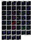
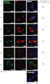
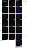
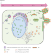
Here, we used a biologically contained Ebola virus system to characterize the spatio-temporal distribution of Ebola virus proteins and RNA during virus replication. We found that viral nucleoprotein (NP), the polymerase cofactor VP35, the major matrix protein VP40, the transcription activator VP30, and the minor matrix protein VP24 were distributed in cytoplasmic inclusions. These inclusions enlarged near the nucleus, became smaller pieces, and subsequently localized near the plasma membrane. GP was distributed in the cytoplasm and transported to the plasma membrane independent of the other viral proteins. We also found that viral RNA synthesis occurred within the inclusions. Newly synthesized negative-sense RNA was distributed inside the inclusions, whereas positive-sense RNA was distributed both inside and outside. These findings provide useful insights into Ebola virus replication.
Figures


References
-
- Sanchez A., Geisbert T. & Feldmann H. Filoviridae: Marburg and Ebola viruses, p 1409–1448. In Knipe, D. M., Howley, P. M. (ed), Fields virology, 5th ed. Lippincott/Williams & Wilkins Co, Philadelphia, PA. (2007).
-
- Becker S., Rinne C., Hofsass U., Klenk H. D. & Muhlberger E. Interactions of Marburg virus nucleocapsid proteins. Virology 249 (2), 406–417 (1998). - PubMed
Publication types
MeSH terms
Substances
Grants and funding
LinkOut - more resources
Full Text Sources
Other Literature Sources
Medical
Miscellaneous

