

Relationship of neighboring tissue and gliosis to α-synuclein pathology in a fetal transplant for Parkinson's disease
- PMID: 23383381
- PMCID: PMC3560449
Relationship of neighboring tissue and gliosis to α-synuclein pathology in a fetal transplant for Parkinson's disease
Abstract
Background: Fetal transplantation for Parkinson disease (PD) had been considered a promising therapeutic strategy; however, reports of Lewy bodies (LBs) and Lewy neurites (LNs) in engrafted tissue adds to controversy surrounding this treatment for PD.
Methods: The brain of a PD patient who had fetal transplantation 14 years before death was evaluated. The graft was studied with routine histologic methods, as well as immunohistochemistry for α-synuclein, neurofilament, synaptophysin and tyrosine hydroxylase (TH), as well as glial fibrillary acidic protein (GFAP) for astrocytes and ionized calcium-binding adaptor molecule 1 (IBA-1) for microglia.
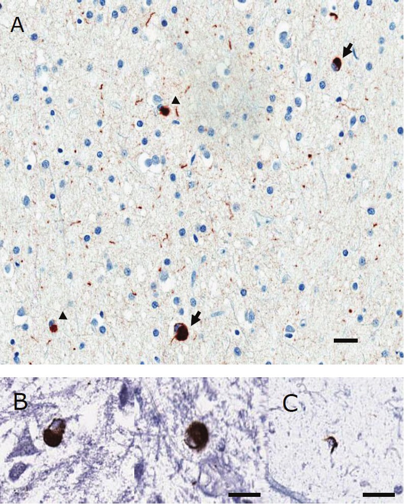
Results: On coronal sections of the brain, the graft extended from the putamen to the amygdala, abutting the anterior hippocampus. Microscopically, the graft consisted of neuron-rich and glia-rich portions. Neuron-rich portions, resembling a neuronal heterotopia, were located in the putamen, whereas the glia-rich portion was more ventral near the amygdala. LBs and LNs were detected in the ventral portion of the graft, especially that part of the graft within the amygdala. Areas with LBs and LNs also had astrogliosis and microgliosis. TH positive neurons were rare and their distribution did not overlap with LBs or LNs.
Comments: LBs and LNs were detected in the transplanted tissue with α-synuclein immunohistochemistry. Unexpected outgrowth of the graft into the amygdala was accompanied by skewed distribution of LBs and gliosis, more abundant in the graft within the amygdala. The distribution of LBs within the graft may suggest the potential role of the local environment as well as gliosis in formation of α-synuclein pathology.
Keywords: Fetal transplantation; Parkinson disease (PD); gliosis; therapy; α-synuclein pathology.
Figures


References
-
- Olanow CW, Goetz CG, Kordower JH, Stoessl AJ, Sossi V, Brin MF, Shannon KM, Nauert GM, Perl DP, Godbold J, Freeman TB. A double-blind controlled trial of bilateral fetal nigral transplantation in Parkinson's disease. Ann Neurol. 2003;54:403–414. - PubMed
-
- Freed CR, Greene PE, Breeze RE, Tsai WY, DuMouchel W, Kao R, Dillon S, Winfield H, Culver S, Trojanowski JQ, Eidelberg D, Fahn S. Transplantation of embryonic dopamine neurons for severe Parkinson's disease. N Engl J Med. 2001;344:710–719. - PubMed
-
- Kordower JH, Chu Y, Hauser RA, Freeman TB, Olanow CW. Lewy body-like pathology in long-term embryonic nigral transplants in Parkinson's disease. Nat Med. 2008;14:504–506. - PubMed
-
- Kordower JH, Chu Y, Hauser RA, Olanow CW, Freeman TB. Transplanted dopaminergic neurons develop PD pathologic changes: a second case report. Mov Disord. 2008;23:2303–2306. - PubMed
-
- Li JY, Englund E, Holton JL, Soulet D, Hagell P, Lees AJ, Lashley T, Quinn NP, Rehncrona S, Bjorklund A, Widner H, Revesz T, Lindvall O, Brundin P. Lewy bodies in grafted neurons in subjects with Parkinson's disease suggest host-to-graft disease propagation. Nat Med. 2008;14:501–503. - PubMed
Grants and funding
LinkOut - more resources
Full Text Sources
Miscellaneous