. 2013 Mar 5;52(9):1611-21.
doi: 10.1021/bi301702p. Epub 2013 Feb 21.

The Qdot-labeled actin super-resolution motility assay measures low-duty cycle muscle myosin step size

Affiliations

The Qdot-labeled actin super-resolution motility assay measures low-duty cycle muscle myosin step size

Yihua Wang et al. Biochemistry. .

Abstract

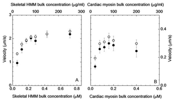
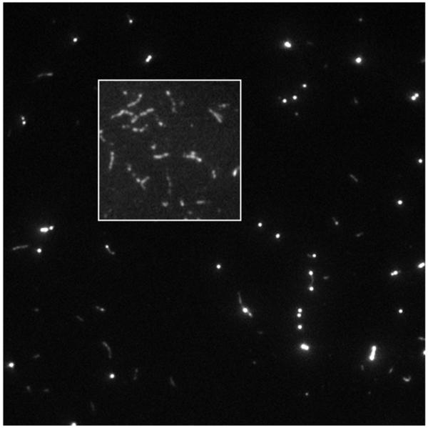
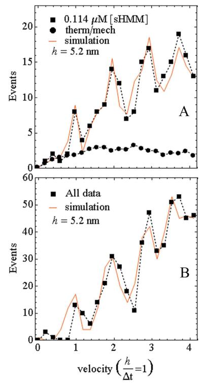
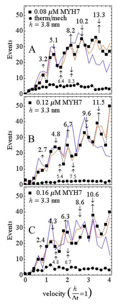
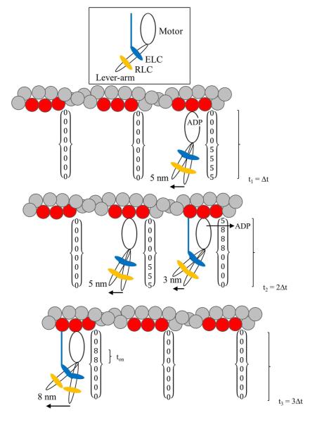
Myosin powers contraction in heart and skeletal muscle and is a leading target for mutations implicated in inheritable muscle diseases. During contraction, myosin transduces ATP free energy into the work of muscle shortening against resisting force. Muscle shortening involves relative sliding of myosin and actin filaments. Skeletal actin filaments were fluorescently labeled with a streptavidin conjugate quantum dot (Qdot) binding biotin-phalloidin on actin. Single Qdots were imaged in time with total internal reflection fluorescence microscopy and then spatially localized to 1-3 nm using a super-resolution algorithm as they translated with actin over a surface coated with skeletal heavy meromyosin (sHMM) or full-length β-cardiac myosin (MYH7). The average Qdot-actin velocity matches measurements with rhodamine-phalloidin-labeled actin. The sHMM Qdot-actin velocity histogram contains low-velocity events corresponding to actin translation in quantized steps of ~5 nm. The MYH7 velocity histogram has quantized steps at 3 and 8 nm in addition to 5 nm and larger compliance compared to that of sHMM depending on the MYH7 surface concentration. Low-duty cycle skeletal and cardiac myosin present challenges for a single-molecule assay because actomyosin dissociates quickly and the freely moving element diffuses away. The in vitro motility assay has modestly more actomyosin interactions, and methylcellulose inhibited diffusion to sustain the complex while preserving a subset of encounters that do not overlap in time on a single actin filament. A single myosin step is isolated in time and space and then characterized using super-resolution. The approach provides a quick, quantitative, and inexpensive step size measurement for low-duty cycle muscle myosin.


Figures

Figure 1
Figure 1
Ensemble average in vitro motility velocity vs bulk concentration of myosin for the rhodamine-phalloidin (open circles) and Qdot+rhodamine-phalloidin (closed circles) labeled actin. Error bars show standard deviation. (A) sHMM and (B) MYH7.
Figure 2
Figure 2
Qdot+rhodamine-phalloidin labeled actin filaments bound to sHMM in the motility assay. The inset shows rhodamine labeled actin filaments in the motility assay with rhodamine-phalloidin to phalloidin in a molar ratio 1:9. Rhodamine labeling is heterogeneous along the filament at this labeling density
Figure 3
Figure 3
The high contrast, intensity-inverted, Qdot track in an in vitro motility assay of sHMM. The QuickPALM super-resolved single particles are rendered at one pixel resolution and all previously recorded frames are plotted in the image (Type 2 rendering). Circles connected by a line are the MTrackJ manually created track. The circle is placed manually within a few pixels of the super-resolved particle position. The track is then associated to the correct super-resolved particle using another computer program, SRTrack, described in the text.
Figure 4
Figure 4
Actin sliding velocity event distribution for 50 msec frame capture intervals. (A) Event distribution for the 0.114 μM bulk sHMM concentration (closed squares connected by dashed line) and the simulation (solid line). The fitted baseline representing thermal/mechanical velocity fluctuation (closed circles connected by dashed line) is also shown. (B) Same as in A except for the event distribution summed over all bulk concentrations. Simulated lines are generated as described in the text.
Figure 5
Figure 5
Actin sliding velocity event distribution for 200 msec frame capture intervals. Single vertical arrows identify unitary steps (↼ short, ⇑ medium, ⒑ long), and their correlation to peaks in the velocity histogram. Arrow orientation (up or down) is for clarity and has no significance. Combined vertical arrows indicate multiple unitary step combinations. Numbers above the vertical arrows indicate observed steps in nm computed by multiplying the ordinate position with the step parameter, h. Smaller font step lengths below the curves are two short or one short-one medium combinations that are often not evidentially present in the experimental data. (A) Event distribution for the 0.08 μM bulk MYH7 concentration (closed squares connected by dashed line) and simulations (solid lines). Simulations include 2 (blue) or 3 (red) unitary steps. Fitting parameter h = 3.8. The fitted baseline (closed circles connected by dashed line) represents thermal/mechanical velocity fluctuation. (B and C) Same as in A except for 0.12 and 0.16 μM bulk MYH7 concentration and fitting parameter h = 3.3.
Figure 6
Figure 6
The three steps of MYH7 and their representation in the time×space array used in the simulation. MYH7 cross-bridge has a motor domain, lever-arm, and two light chains ELC (blue) and RLC (orange) indicated at the top. The actin/ELC linkage, shown as a line connecting ELC with actin, modulates myosin step size. The 3 actin monomers in the target zone are shown in red. Each target zone has a column in the time×space array. The figure shows three frame capture intervals, Δt, with frame capture happening at t = t1, t2, and t3. The cross-bridge attached at the upper right performs the major 5 nm step giving the 5 nm displacement recorded at t1. Slow ADP release is indicated in the 5 nm step cycle shown. This cross-bridge remains attached into the second exposure interval when it releases ADP then converts to the 8 nm step with the ELC linkage and produces the minor 3 nm displacement. Additional displacement of 5 nm by the middle cross-bridge releases ADP without the ELC linkage in the second exposure interval giving 3+5 nm total displacement at t2 due to two different unitary steps. The third exposure interval has a unitary 8 nm displacement caused by a minor step from a single cross-bridge.

References

    1. Moore JR, Leinwand L, Warshaw DM. Understanding Cardiomyopathy Phenotypes Based on the Functional Impact of Mutations in the Myosin Motor. Circ. Res. 2012;111:375–385. - PMC - PubMed
    1. Tajsharghi H, Kimber E, Kroksmark AK, Jerre R, Tulinius M, Oldfors A. Embryonic Myosin Heavy-Chain Mutations Cause Distal Arthrogryposis and Developmental Myosin Myopathy That Persists Postnatally. Arch.Neurology. 2008;65:1083–1090. - PubMed
    1. Rayment I, Rypniewski WR, Schmidt-Base K, Smith R, Tomchick DR, Benning MM, Winkelmann DA, Wesenberg G, Holden HM. Three-dimensional structure of myosin subfragment-1: A molecular motor. Science. 1993;261:50–58. - PubMed
    1. Dominguez R, Freyzon Y, Trybus KM, Cohen C. Crystal structure of a vertebrate smooth muscle myosin motor domain and its complex with the essential light chain: visualization of the pre-power stroke state. Cell. 1998;94:559–571. - PubMed
    1. Sherwood JJ, Waller GS, Warshaw DM, Lowey S. A point mutation in the regulatory light chain reduces the step size of skeletal muscle myosin. Proceedings of the National Academy of Sciences. 2004;101:10973–10978. - PMC - PubMed

Publication types

MeSH terms

LinkOut - more resources