. 2013 Feb 6;14(1):15.
doi: 10.1186/1465-9921-14-15.

Novel immune genes associated with excessive inflammatory and antiviral responses to rhinovirus in COPD

Affiliations

Novel immune genes associated with excessive inflammatory and antiviral responses to rhinovirus in COPD

Katherine J Baines et al. Respir Res. .

Abstract

Background: Rhinovirus (RV) is a major cause of chronic obstructive pulmonary disease (COPD) exacerbations, and primarily infects bronchial epithelial cells. Immune responses from BECs to RV infection are critical in limiting viral replication, and remain unclear in COPD. The objective of this study is to investigate innate immune responses to RV infection in COPD primary BECs (pBECs) in comparison to healthy controls.

Methods: Primary bronchial epithelial cells (pBECs) from subjects with COPD and healthy controls were infected with RV-1B. Cells and cell supernatant were collected and analysed using gene expression microarray, qPCR, ELISA, flow cytometry and titration assay for viral replication.

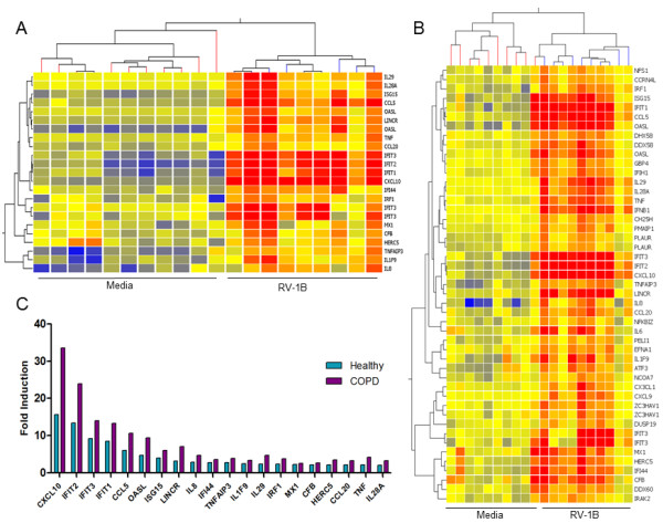
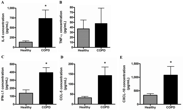
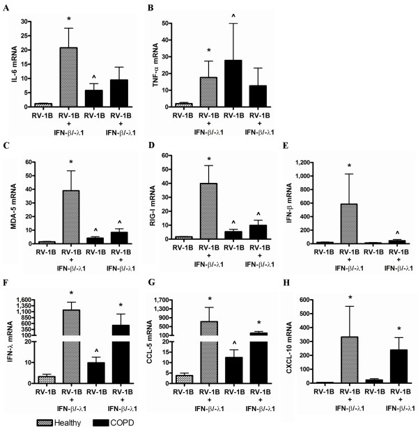
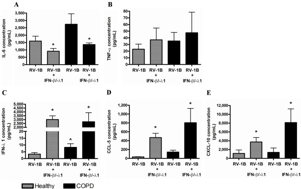
Results: COPD pBECs responded to RV-1B infection with an increased expression of antiviral and pro-inflammatory genes compared to healthy pBECs, including cytokines, chemokines, RNA helicases, and interferons (IFNs). Similar levels of viral replication were observed in both disease groups; however COPD pBECs were highly susceptible to apoptosis. COPD pBECs differed at baseline in the expression of 9 genes, including calgranulins S100A8/A9, and 22 genes after RV-1B infection including the signalling proteins pellino-1 and interleukin-1 receptor associated kinase 2. In COPD, IFN-β/λ1 pre-treatment did not change MDA-5/RIG-I and IFN-β expression, but resulted in higher levels IFN-λ1, CXCL-10 and CCL-5. This led to reduced viral replication, but did not increase pro-inflammatory cytokines.

Conclusions: COPD pBECs elicit an exaggerated pro-inflammatory and antiviral response to RV-1B infection, without changing viral replication. IFN pre-treatment reduced viral replication. This study identified novel genes and pathways involved in potentiating the inflammatory response to RV in COPD.


Figures

Figure 1
Figure 1
Cluster analysis of genes significantly induced in healthy (n=10) and COPD (n=10) pBECs 6 hours after RV-1B infection. Microarray analysis showed that RV-1B infection significantly up-regulated (A) 20 genes in healthy controls and (B) 42 genes in COPD pBECs. (C) All the genes that were up-regulated in both groups were significantly higher in COPD compared to that in healthy control pBECs.
Figure 2
Figure 2
Inflammatory and antiviral genes at 6 hours after RV-1B infection in healthy (n=10) and COPD (n=10) pBECs, confirmation of the microarray findings. Inflammatory cytokines (A) IL-6 and (B) TNF-α were not induced by RV-1B infection in healthy control pBECs, but were IL-6 was significantly up-regulated by infection in COPD pBECs, whilst TNF-α was significantly higher in the baseline media control and after RV infection. RNA helicases (C) MDA-5 and (D) RIG-I and antiviral (E) IFN-β, (F) IFN-λ1, (G) CCL-5 and (H) CXCL-10 mRNA were all significantly higher in RV infected COPD pBECs compared to healthy controls. Results are presented as mean fold change in expression with the error bar as standard error of the mean (SEM). * p<0.05 versus the corresponding media control. ^ p<0.05 versus healthy RV infected pBECs.
Figure 3
Figure 3
Inflammatory and antiviral proteins at 24 hours after RV-1B infection in healthy (n=10) and COPD (n=10) pBECs. (A) IL-6 protein induction after infection was significantly higher in COPD pBECs however (B) TNF-α induction was not significantly different between healthy and COPD pBECs. (C) IFN-λ1, (D) CCL-5 and (E) CXCL-10 protein induction in COPD pBECs was also significantly higher than that in healthy pBECs. Results are presented as mean and the error bar as standard error of the mean (SEM). * p<0.05 versus healthy RV infected pBECs.
Figure 4
Figure 4
Key signalling molecules and pathways involved in RV induced inflammatory responses in COPD pBECs. Genes that were identified to play a role in innate immune signalling that were increased in RV infected COPD pBECs include IRAK2, PELI1, CH25H, PMAIP1, ATF3 and GBP4. IRAK2, PELI1 and CH25H (bolded in box) are involved in signal transduction downstream of IL-1/TLR leading to NF-κB activation and production of inflammatory cytokines. Induction of PMAIP1, a mitochondria-associated protein, leads to apoptosis. ATF3 senses cellular stress and functions to reduce TLR4-mediated NF-κB signalling. GBP4 is an IFN-inducible GTPase that has been shown to disrupt IRF7 activation, thereby inhibiting the induction of type I IFNs. Solid line indicates up-regulation, and dashed line indicates down-regulation.
Figure 5
Figure 5
Inflammatory and antiviral genes at 6 hours after RV-1B infection in IFN-β/λ1-pre-treated healthy (n=6) and COPD (n=6) pBECs. IFN-β/λ1-pre-treatment before RV-1B infection resulted in a significant increase in (A) IL-6 and (B) TNF-α mRNA in healthy but not in COPD pBECs. (C) MDA-5, (D) RIG-I, and (E) IFN-β mRNA was significantly induced and higher in healthy control than in COPD pBECs. In sharp contrast, (F) IFN-λ1, (G) CCL-5, and (H) CXCL-10 mRNA was significantly up-regulated by IFNs pre-treatment in both healthy and COPD pBECs. Results were presented as mean fold change in expression with and the error bar as standard error of the mean (SEM). * p<0.05 versus the corresponding RV-1B alone. ^p<0.05 versus healthy RV-1B infected pBECs.
Figure 6
Figure 6
Inflammatory and antiviral proteins at 24 hours after RV-1B infection in IFN-β/λ1-pre-treated healthy (n=6) and COPD (n=6) pBECs. IFN-β/λ1-pre-treatment before RV-1B infection led to a reduction of (A) IL-6 in healthy pBECs, but had no effect in COPD pBECs. (B) Similarly TNF-α protein was not affected by IFNs pre-treatment. (C) IFN-λ1, (D) CCL-5, and (E) CXCL-10 was significantly induced in pre-treated healthy and COPD pBECs. Results were presented as standard error of the mean (SEM). * p<0.05 versus the corresponding RV-1B. ^p<0.05 versus healthy RV-1B infected pBECs.

References

    1. MacNee W. Pathogenesis of Chronic Obstructive Pulmonary Disease. Proc Am Thor Soc. 2005;2:258–266. doi: 10.1513/pats.200504-045SR. - DOI - PMC - PubMed
    1. Mallia P, Message SD, Kebadze T, Parker HL, Kon OM, Johnston SL. An experimental model of rhinovirus induced chronic obstructive pulmonary disease exacerbations: a pilot study. Respir Res. 2006;7:116. doi: 10.1186/1465-9921-7-116. - DOI - PMC - PubMed
    1. Mosser AG, Brockman-Schneider R, Amineva S, Burchell L, Sedgwick JB, Busse WW, Gern JE. Similar Frequency of Rhinovirus-Infectible Cells in Upper and Lower Airway Epithelium. J Infect Dis. 2002;185:734–743. doi: 10.1086/339339. - DOI - PubMed
    1. Gern JE, Dick EC, Lee WM, Murray S, Meyer K, Handzel ZT, Busse WW. Rhinovirus enters but does not replicate inside monocytes and airway macrophages. J Immunol. 1996;156:621–627. - PubMed
    1. Seth RB, Sun L, Chen ZJ. Antiviral innate immunity pathways. Cell Res. 2006;16:141–147. doi: 10.1038/sj.cr.7310019. - DOI - PubMed

MeSH terms