. 2013 Feb 6:13:27.
doi: 10.1186/1471-2180-13-27.

Transcriptional analysis of ftsZ within the dcw cluster in Bacillus mycoides

Affiliations

Transcriptional analysis of ftsZ within the dcw cluster in Bacillus mycoides

Tiziana Santini et al. BMC Microbiol. .

Abstract

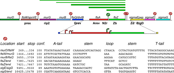
Background: In Bacillus mycoides, as well as in other members of the B. cereus group, the tubulin-like protein of the division septum FtsZ is encoded by the distal gene of the cluster division and cell wall (dcw). Along the cluster the genes coding for structural proteins of the division apparatus are intermingled with those coding for enzymes of peptidoglycan biosynthesis, raising the possibility that genes with this different function might be coexpressed. Transcription of ftsZ in two model bacteria had been reported to differ: in B. subtilis, the ftsZ gene was found transcribed as a bigenic mRNA in the AZ operon; in E. coli, the transcripts of ftsZ were monogenic, expressed by specific promoters. Here we analyzed the size and the initiation sites of RNAs transcribed from ftsZ and from other cluster genes in two B. mycoides strains, DX and SIN, characterized by colonies of different chirality and density, to explore the correlation of the different morphotypes with transcription of the dcw genes.

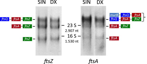
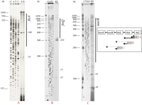
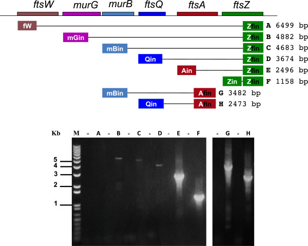
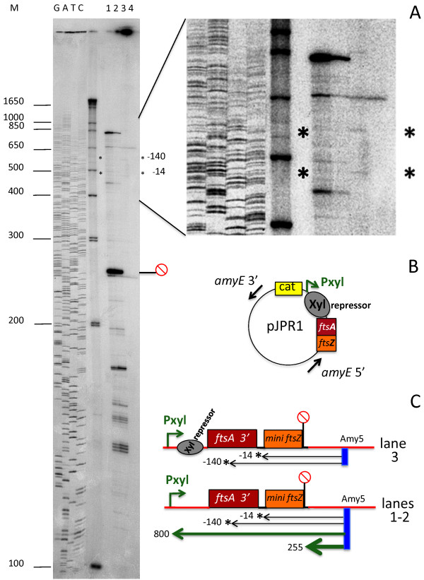
Results: In both strains, during vegetative growth, the ftsZ-specific RNAs were composed mainly of ftsZ, ftsA-ftsZ and ftsQ-ftsA-ftsZ transcripts. A low number of RNA molecules included the sequences of the upstream murG and murB genes, which are involved in peptidoglycan synthesis. No cotranscription was detected between ftsZ and the downstream genes of the SpoIIG cluster. The monogenic ftsZ RNA was found in both strains, with the main initiation site located inside the ftsA coding sequence. To confirm the promoter property of the site, a B. mycoides construct carrying the ftsA region in front of the shortened ftsZ gene was inserted into the AmyE locus of B. subtilis 168. The promoter site in the ftsA region was recognized in the heterologous cellular context and expressed as in B. mycoides.

Conclusions: The DX and SIN strains of B. mycoides display very similar RNA transcription specificity. The ftsZ messenger RNA can be found either as an independent transcript or expressed together with ftsA and ftsQ and, in low amounts, with genes that are specific to peptidoglycan biosynthesis.


Figures

Figure 1
Figure 1
Northern blot analysis of RNA from exponentially growing B. mycoides SIN and DX. SIN and DX total RNA was electrophoresed in formaldehyde-agarose and blotted. The same filter was hybridized first to ftsZ and, after stripping, to ftsA DNA probes. The position of ribosomal 23S (2907 bases) and 16S (1530 bases) RNA on the filter is indicated. FtsZ and ftsA RNAs in the band below 16S rRNA are monogenic transcripts. The band below the position of the 23 S rRNA contains the ftsA-ftsZ bigenic transcripts. The transcripts of the genes ftsQ-ftsA-ftsZ are within the uppermost bands together with the transcripts murB-ftsQ-ftsA, detected only by the ftsA probe.
Figure 2
Figure 2
Determination of ftsZ, ftsA and ftsQ RNA 5’ ends by primer extension (PE) in B. mycoides SIN (S) and DX (D). 5’ 32P-labeled primers were hybridized to total RNA, extended by reverse transcriptase and the cDNAs separated by 6% urea-PAGE electrophoresis. The numbers on the right side of the autoradiograms indicate the position of the cDNA 3’ ends relative to the ORF first nucleotide (+1). The thick lateral bar indicates the approximate position in the gel of the next upstream gene. A) PE from primer ZB annealing to ftsZ RNA at +103; B) PE from primer Arev annealing to ftsA RNA at +80; C) PE from primer Qrev annealing to ftsQ RNA at +52 (Table 1). Here two different SIN cDNA preparations were loaded on the gel. A schematic view of the major cDNA products is shown in the inset. M = MW marker 32P-labeled DNAs. GATC = 35S-dATP labeled M13mp18 ladder.
Figure 3
Figure 3
RT-PCR analysis of RNA transcripts from the dcw genes in B. mycoides. Purified vegetative RNA of B. mycoides DX was reverse transcribed from primers complementary to the 3’ end of ftsZ (Zfin) and to the 3’ end of ftsA (Afin). The control cDNAs (lanes -) were without RT in the reaction. cDNAs were PCR amplified using Zfin (A-F) and Afin (G-H) as downstream primers. Upstream primers were specific for each gene (Table 1). Multigene ftsZ RNAs included murG and murB, though not ftsW transcripts.
Figure 4
Figure 4
Initiation of mini-ftsZ RNA transcripts in B. subtilis. The B. mycoides mini-ftsZ DNA construct was cloned into pJPR1 and inserted at the AmyE site of B. subtilis 168 (see methods). Transcripts of the construct were detected in total B. subtilis RNA by primer extension from the labeled primer Amy5 (Table 1) specific to the amyE 5’ region located 245 nt downstream of the inserted construct. A) Autoradiogram of PE. Lanes1 and 2: transcripts originating from the Pxyl promoter, induced by 5% xylose for 18 and 3 hours. Lane 3: the faint transcripts of the ftsZ minigene present in the non-induced B. subtilis recombinant strain are indicated by asterisks and map at −140 and −10 from the first nucleotide of the minigene ftsZ ORF as in B. mycoides. These bands are not present in the control B. subtilis strain (lane 4). B) schematic view of the construct in pJPR1. C) Schematic representation of the cDNAs indicated by asterisks in A. The red circle marks the position of the terminator structure 3’ to the B. mycoides ftsZ ORF. M = MW marker DNA. GATC = M13MP18 sequence ladder.
Figure 5
Figure 5
Transcriptional terminators within the B. mycoides dcw and spoIIG gene clusters. Red labels mark the position of the putative terminators. The DX termination sequences displayed are 100% identical to those predicted at the TransTerm-HP site for B. weihenstephanensis KBAB4 (Accession NC_010184, from coordinates 3780796 to 3790953). The green label between ftsA and ftsZ indicates a hairpin structure not recognized there as a potential terminator. The three large green bars over the genes represent the main ftsZ-specific RNAs and the green thin bars the minor ones. The primers used to detect RNA 5’ ends by primer extension are indicated below the genes. The curved arrows in the enlarged region show the main ftsA and ftsZ RNA start sites.

References

    1. Schmidt TR, Scott EJ II, Dyer DW. Whole-genome phylogenies of the family Bacillaceae and expansion of the sigma factor gene family in the Bacillus cereus species-group. BMC Genomics. 2011;12:430. doi: 10.1186/1471-2164-12-430. - DOI - PMC - PubMed
    1. Gause GF. Some physiological properties of dextral and of sinistral forms in Bacillus mycoides flügge. Biol Bull Woods Hole MA. 1939;76:448–465. doi: 10.2307/1537751. - DOI
    1. Di Franco C, Beccari E, Santini T, Pisaneschi G, Tecce G. Colony shape as a genetic trait in the pattern-forming Bacillus mycoides. BMC Microbiol. 2002;2(33):1–15. - PMC - PubMed
    1. Turchi L, Santini T, Beccari E, Di Franco C. Localization of new peptidoglycan at poles in Bacillus mycoides, a member of the Bacillus cereus group. Arch Microbiol. 2012;194(10):887–892. doi: 10.1007/s00203-012-0830-1. - DOI - PMC - PubMed
    1. Gholamhoseinian A, Shen Z, Wu J-J, Piggot P. Regulation of transcription of the cell division gene ftsA during sporulation of Bacillus subtilis. J Bacteriol. 1992;174(14):4647–4656. - PMC - PubMed

Publication types

MeSH terms

Associated data

LinkOut - more resources