. 2013 Feb 6:13:5.
doi: 10.1186/1472-6890-13-5.

A simple immunohistochemical panel comprising 2 conventional markers, Ki67 and p53, is a powerful tool for predicting patient outcome in luminal-type breast cancer

Affiliations

A simple immunohistochemical panel comprising 2 conventional markers, Ki67 and p53, is a powerful tool for predicting patient outcome in luminal-type breast cancer

Takayuki Kobayashi et al. BMC Clin Pathol. .

Abstract

Background: Ki67 is widely used in order to distinguish the "A" and "B" subtypes of luminal-type breast cancer. This study aimed to validate the prognostic value of adding p53 to Ki67 for characterizing luminal-type breast cancer.

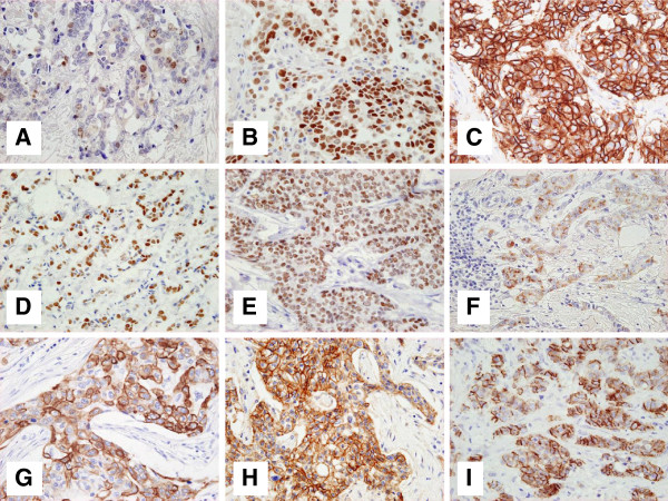
Methods: Immunostaining for Ki67, p53, and the molecular markers HER2, CK5/6, CK14, EGFR, FOXA1, GATA3, and P-cadherin was examined hormone receptor (HR)-positive cancer tissues from 150 patients. The prognostic value of an immunohistochemical panel comprising Ki67 and p53 was compared with that of the single Ki67 labeling index (LI), and uni- and multivariate analyses were performed.

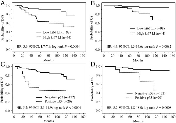
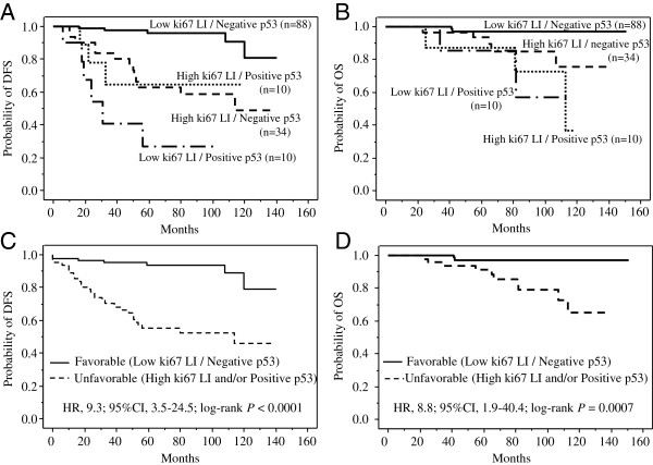
Results: Division of the patients based on the immunohistochemistry results into favorable- (low Ki67 LI, p53-negative) and unfavorable- (high Ki67 LI and/or p53-positive) phenotype groups yielded distinctly different Kaplan-Meier's curves of both disease-free (P<0.0001) and overall survival (P=0.0007). These differences were much more distinct than those between the corresponding low Ki67 LI vs. high Ki67LI curves. While the prognostic values of the other molecular markers were not significant, combined Ki67-p53 status was an independent prognostic factor by multivariate analysis.

Conclusion: These data indicate that an immunohistochemical panel comprising Ki67 and p53 is a practical tool for management of patients with HR-positive breast cancer.


Figures

Figure 1
Figure 1
Representative images of immunostaining for 9 molecular markers.A, Positive nuclear Ki67 staining. The Ki67 labeling index of this specimen is 12%. B, p53 staining in the nucleus. This tumor was scored as p53-positive. C, Positive HER2 membrane staining. The HER2 expression of this tumor was scored as 3+. D, Positive nuclear FOXA1 staining. This tumor was classified as FOXA1-positive. E, Positive nuclear GATA3 staining. This tumor was classified as GATA3-positive. F, Positive CK5/6 membrane or sub-membrane staining of tumor cells. This tumor was classified as CK5/6-positive. G, Positive CK14 membrane or sub-membrane staining of tumor cells. This tumor was classified as CK14-positive. H, Positive EGFR membrane staining of tumor cells. The EGFR expression of this tumor was scored as 3+. I, Positive P-cadherin membrane or sub-membrane immunoreactivity of tumor cells. This tumor was classified as P-cadherin-high. The magnification of all figures is ×400.
Figure 2
Figure 2
Prognostic impact of Ki67 labeling index (LI) and p53 status in patients with hormone receptor-positive and HER2-negative breast cancer (n=142).A, Disease free survival curves for 98 patients with Ki67 LI-low tumors and 44 patients with Ki67 LI-high tumors. The 2 curves differ significantly (HR, 3.6; 95% CI, 1.7–7.9; log-rank P = 0.0004). B, Overall survival curves for 98 patients with Ki67 LI-low tumors and 44 patients with Ki67 LI-high tumors. The 2 curves differ significantly (HR, 4.4; 95% CI, 1.3–14.6; log-rank P = 0.0082). C, Disease-free survival curves for 122 patients with p53-negative tumors and 20 patients with p53-positive tumors. The 2 curves differ significantly (HR, 5.2; 95% CI, 2.3–11.9; log-rank P < 0.0001). D, Overall survival curves for 122 patients with p53-negative tumors and 20 patients with p53-positive tumors. The 2 curves differ significantly (HR, 5.7; 95% CI, 1.8–18.0; log-rank P = 0.0008).
Figure 3
Figure 3
Prognostic impact of combined Ki67-p53 status in patients with hormone receptor-positive and HER2-negative breast cancer (n=142). A, Disease-free survival curves for 88 patients with Ki67 LI-low and p53-negative tumors, 34 patients with Ki67 LI-high and p53-negative tumors, 10 patients with Ki67 LI-low and p53-positive tumors, and 10 patients with Ki67 LI-high and p53-positive tumors. Patients with Ki67 LI-low and p53-negative tumors had significantly longer disease-free survival than those with Ki67 LI-low and p53-positive tumors, those with Ki67 LI-high and p53-negative tumors, or those with Ki67 LI-high and p53-positive tumors (P < 0.0001, P < 0.0001, and P = 0.0005, respectively). B, Overall survival curves for 88 patients with Ki67 LI-low and p53-negative tumors, 34 patients with Ki67 LI-high and p53-negative tumors, 10 patients with Ki67 LI-low and p53-positive tumors, and 10 patients with Ki67 LI-high and p53-positive tumors. Patients with Ki67 LI-low and p53-negative tumors had significantly longer disease-free survival than those with Ki67 LI-low and p53-positive tumors, those with Ki67 LI-high and p53-negative tumors, or those with Ki67 LI-high and p53-positive tumors (P = 0.0010, P = 0.011, and P < 0.0001, respectively). C, Disease-free survival curves for patients with favorable-phenotype tumors (88 patients with Ki67 LI-low and p53-negative tumors) and unfavorable-phenotype tumors (54 patients with Ki67 LI-high and/or p53-positive tumors). The disease-free survival time was significantly longer in the favorable-phenotype group than in the unfavorable-phenotype group (HR, 9.3; 95% CI, 3.5–24.5; P < 0.0001). D, Overall survival curves for patients with favorable-phenotype tumors (88 patients with Ki67 LI-low and p53-negative tumors) and those with unfavorable-phenotype tumors (54 patients with Ki67 LI-high and/or p53-positive tumors). The disease-free survival was significantly longer in the favorable-phenotype group than in the unfavorable-phenotype group (HR, 8.8; 95% CI, 1.9–40.4; P = 0.0007).

References

    1. Badve S, Nakshatri H. Oestrogen-receptor-positive breast cancer: towards bridging histopathological and molecular classifications. J Clin Pathol. 2009;62(1):6–12. doi: 10.1136/jcp.2008.059899. - DOI - PubMed
    1. Hortobagyi GN. Treatment of breast cancer. N Engl J Med. 1998;339(14):974–984. doi: 10.1056/NEJM199810013391407. - DOI - PubMed
    1. Early Breast Cancer Trialists' Collaborative Group. Tamoxifen for early breast cancer: an overview of the randomised trials. Lancet. 1998;351(9114):1451–1467. - PubMed
    1. Mauri D, Pavlidis N, Polyzos NP, Ioannidis JP. Survival with aromatase inhibitors and inactivators versus standard hormonal therapy in advanced breast cancer: meta-analysis. J Natl Cancer Inst. 2006;98(18):1285–1291. doi: 10.1093/jnci/djj357. - DOI - PubMed
    1. Fisher B, Jeong JH, Bryant J, Anderson S, Dignam J, Fisher ER, Wolmark N. Treatment of lymph-node-negative, oestrogen-receptor-positive breast cancer: long-term findings from National Surgical Adjuvant Breast and Bowel Project randomised clinical trials. Lancet. 2004;364(9437):858–868. doi: 10.1016/S0140-6736(04)16981-X. - DOI - PubMed