Review
. 2013:81:27-76.
doi: 10.1016/B978-0-12-407826-0.00002-3.

Red blood cell polymorphism and susceptibility to Plasmodium vivax

Affiliations
Review

Red blood cell polymorphism and susceptibility to Plasmodium vivax

Peter A Zimmerman et al. Adv Parasitol. 2013.

Abstract

Resistance to Plasmodium vivax blood-stage infection has been widely recognised to result from absence of the Duffy (Fy) blood group from the surface of red blood cells (RBCs) in individuals of African descent. Interestingly, recent studies from different malaria-endemic regions have begun to reveal new perspectives on the association between Duffy gene polymorphism and P. vivax malaria. In Papua New Guinea and the Americas, heterozygous carriers of a Duffy-negative allele are less susceptible to P. vivax infection than Duffy-positive homozygotes. In Brazil, studies show that the Fy(a) antigen, compared to Fy(b), is associated with lower binding to the P. vivax Duffy-binding protein and reduced susceptibility to vivax malaria. Additionally, it is interesting that numerous studies have now shown that P. vivax can infect RBCs and cause clinical disease in Duffy-negative people. This suggests that the relationship between P. vivax and the Duffy antigen is more complex than customarily described. Evidence of P. vivax Duffy-independent red cell invasion indicates that the parasite must be evolving alternative red cell invasion pathways. In this chapter, we review the evidence for P. vivax Duffy-dependent and Duffy-independent red cell invasion. We also consider the influence of further host gene polymorphism associated with malaria endemicity on susceptibility to vivax malaria. The interaction between the parasite and the RBC has significant potential to influence the effectiveness of P. vivax-specific vaccines and drug treatments. Ultimately, the relationships between red cell polymorphisms and P. vivax blood-stage infection will influence our estimates on the population at risk and efforts to eliminate vivax malaria.


Figures

Figure 2.1
Figure 2.1. Plasmodium knowlesi invasion of Macaca mulatta red blood cells
A P. knowlesi merozoite has commenced the invasion process through formation of gliding junctions involving merozoite and red cell membranes. This enables invagination of the erythrocyte membrane and movement of the parasite into the parasitophorous vacuole. R, rhoptry; M, micronemes; J, gliding junction; PV, parasitophorous vacuole; E, erythrocyte. (Figure from unpublished data, Hisashi Fujioka)
Figure 2.2
Figure 2.2. The Duffy antigen
The diagram illustrates the primary structure of the 236-amino-acid 36–46-kDa Duffy antigen with seven predicted transmembrane domains and extracellular and intracellular domains. Amino acids comprising the Fy6 and Fy3 antibody-binding domains are marked by brackets. Amino acid sequence polymorphisms are identified at residues 42 (G vs. D; Fya vs. Fyb), 89 (R vs. C; Fyb vs. Fybweak) and 100 (A vs. T) and the two premature termination codons (W vs. X) at residue positions 96 and 134. Glycosylation sites are identified at amino acid residues N16 and N27. Disulfide bonds occurring between C129 (extracellular loop 2) and C195 (extracellular loop 3) and between C51 (amino terminal head) and C276 (extracellular loop 3) are predicted to contribute to further tertiary structure within the cell membrane as depicted in the inset. Amino acids predicted to comprise the P. vivax binding region are identified in red (Chitnis et al., 1996). Duffy Antigen Function – The Duffy antigen receptor for chemokines (DARC) is a ‘silent’ 7-transmembrane receptor. This results from the absence of a DRYLAIV amino acid motif in the second intracellular loop needed to couple with G-proteins that initiate intracellular signalling cascades (Murphy, 1996). Duffy is one of a few chemokine receptors that bind to inflammatory chemokines, categorised by structural features into two different groups, α (amino acid motif -CC-) and β (amino acid motif –CXC-). On erythrocytes, the Duffy antigen is proposed to act as a sink that binds to excess chemokines and limits inflammation (Darbonne et al., 1991). Reciprocally, Duffy binding of chemokines prevents their diffusion into organs and peripheral tissue space and in this way acts as a reservoir of chemokines in the circulating blood (Fukuma et al., 2003). Duffy is also expressed on a variety of non-erythroid cells including venular endothelial cells; in this context recent studies suggest two potential roles for Duffy. On venular endothelial cells, Duffy has been proposed to act as a chemokine interceptor (internalisation receptor) by internalising and scavenging chemokines (Nibbs et al., 2003). Alternatively, Pruenster et al. have shown that Duffy acts to mediate chemokine transcytosis (Pruenster et al., 2009). In their in_vitro system, Duffy-mediated chemokine transcytosis led to apical retention of intact chemokines and leukocyte migration across Duffy-expressing endothelial cell monolayers. How these complex roles of the Duffy antigen are regulated and influence human health remains to be determined. (Originally published in Zimmerman 2004. The enigma of vivax malaria and erythrocyte Duffy-negativity, in: Dronamraju, K.R., (Ed.), Infectious Disease and Host-Pathogen Evolution.Cambridge University Press, New York, pp 141–172.) (Reproduced with permission from Cambridge University Press and Krishna R. Dronamraju). (For interpretation of the references to colour in this figure legend, the reader is referred to the online version of this book.)
Figure 2.3
Figure 2.3
Standard Giemsa-stained thin smear preparations of P. vivax infection and development in human Duffy-negative erythrocytes. Panels A and B originated from a 4-year-old female, genotyped as Duffy negative (FYBES/BES), who presented at the Tsiroanomandidy health center with fever (37.8 °C), headache and sweating without previous anti-malarial treatment. Standard blood smear diagnosis revealed a mixed infection with P. vivax (parasitaemia = 3040 parasitised red blood cells [pRBC]/μl) and P. falciparum (parasitaemia=980 pRBC/μl). PCR-based Plasmodium species diagnosis confirmed the blood smear result; P. malariae and P. ovale were not detected. (A) a P. vivax early-stage trophozoite with condensed chromatin, enlarged erythrocyte volume, Schüffner stippling and irregular ring-shaped cytoplasm. (B) a P. vivax gametocyte – lavender parasite, larger pink chromatin mass and brown pigment scattered throughout the cytoplasm are characteristics of microgametocytes (male). Panel C originated from a 12- year-old Duffy-negative (FYBES/BES) male, who presented at the Miandrivazo health centre with fever (37.5 °C) and shivering without previous anti-malarial treatment. Standard blood smear diagnosis and light microscopy revealed infection with only P. vivax (parasitaemia = 3000 pRBC/μl). PCR-based Plasmodium species diagnosis confirmed this blood smear result; P. falciparum, P. malariae and P. ovale were not detected. The parasite featured shows evidence of a P. vivax gametocyte – large blue parasite, smaller pink chromatin mass and brown pigment scattered throughout the cytoplasm are characteristics of macrogametocytes (female). (Adapted from Ménard et_al, 2010. Plasmodium vivax clinical malaria is commonly observed in Duffy-negative Malagasy people. Proc. Natl. Acad. Sci. U. S. A. 107, 5967–5971). (For interpretation of the references to colour in this figure legend, the reader is referred to the online version of this book.)
Figure 2.4
Figure 2.4
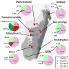
Frequency distribution of P. vivax infections and clinical cases identified in Duffy-positive and Duffy-negative Malagasy people. Pie graphs show the prevalence of Duffy-positive (dark/light green) and Duffy-negative (red/pink quadrants) phenotypes in the eight Madagascar study sites. Prevalence of P. vivax infection observed in the survey of school-aged children is shown in red and dark green; population subsets not infected with P. vivax are pink and light green. Study sites identified by a red star indicate that clinical vivax malaria was observed in Duffy-negative individuals. A green star indicates that vivax malaria was observed in Duffy-positive individuals only (Ejeda). In Ihosy, clinical malaria was observed in one individual with a mixed P. vivax/P. falciparum infection. P. vivax malaria was not observed in Andapa and Farafangana (black star). Malaria transmission strata are identified as tropical (lightest grey), sub-desert (light grey), equatorial (middle grey) and highlands (dark grey). (Adapted from Ménard et_al, 2010. Plasmodium vivax clinical malaria is commonly observed in Duffy-negative Malagasy people. Proc. Natl. Acad. Sci. U. S. A. 107, 5967–5971). (For interpretation of the references to colour in this figure legend, the reader is referred to the online version of this book.)
Figure 2.5
Figure 2.5. Global frequencies of the FY alleles
Areas predominated by a single allele (frequency ≥ 50%) are represented by a colour gradient (blue, FYA; green, FYB; red/yellow, FYBES). Areas of allelic heterogeneity where no single allele predominates, but two or more alleles each have frequencies ≥ 20%, are shown in grey-scale: palest for heterogeneity between the silent FYBES allele and either FYA or FYB (when co-inherited, these do not generate new phenotypes), and darkest being co-occurrence of all three alleles (and correspondingly the greatest genotypic and phenotypic diversity). Overall percentage surface area of each class is listed in the legend. The probability distribution based on a Bayesian model is summarised as a single statistic: in this case, the median value, as this corresponds best to the input dataset, as previously described (Howes et al., 2011). Median values of the predictions were generated for each allele frequency at a 10 × 10 km resolution on a global grid with GIS software (ArcMap 9.3; ESRI). (For interpretation of the references to colour in this figure legend, the reader is referred to the online version of this book.)
Figure 2.6
Figure 2.6. Estimated change in the P. vivax population at risk before (A) and after (B) relaxing the level of resistance conferred by Duffy negativity from 100% to 50%, respectively
Overall increases in the percentage of PvPAR (C). The population-at-risk exclusions are based on the methods developed by Guerra et al. in 2010 (Guerra et al., 2010) and subsequently refined by Gething et al. To define the P. vivax population at risk (PvPAR), Gething et al. imposed several layers of exclusion from the countries with endemic P. vivax transmission (95 countries). Annual parasite incidence (API) data were used to refine the sub-national levels of transmission along administrative boundaries, classifying areas into unstable (<0.1 cases per 1000 population per year), stable transmission (≥0.1 cases per 1000 population per year) and malaria free (Guerra et al., 2010). Temperature exclusion based on minimum requirements for parasite sporogony modelled in relation to vector lifespan (Gething et al., 2011). Aridity mask to exclude areas too dry to sustain transmission by restricting vector survival and availability of ovipositioning sites. The aridity mask was derived from the bare ground areas in the GlobCover land cover imagery (Guerra et al., 2008). Medical intelligence was used to further exclude malaria-free urban areas (modulated with knowledge of the local Anopheles vectors) (Guerra et al., 2010). All these methods have been described in greater detail by Gething et al. (2012) and in Chapter 1 in Volume 80 of this special issue. Extensive data collection was necessary for the API exclusions, and individuals who contributed data to this process are acknowledged on the Malaria Atlas Project website (MAP: www.map.ox.ac.uk/). (For a colour version of this figure, the reader is referred to the online version of this book.)
Figure 2.7
Figure 2.7
Summary of major events providing insight on resistance to P. vivax. (For a colour version of this figure, the reader is referred to the online version of this book.)
Figure 2.8
Figure 2.8. Overview of P. vivax merozoite interaction with the human red blood cell
Initial attachment occurs between any part of the merozoite (blue) and erythrocyte (red). The merozoite reorients, positioning its apical end for attachment to the red cell membrane. A junction forms between the apical end of the merozoite and the erythrocyte membrane of Duffy-positive cells (first call-out box). In contrast, P. knowlesi electron microscopy has shown thin filaments between the merozoite apical end and the Duffy-negative red cell membrane; however, the merozoite is not drawn into contact with the red cell and the junction fails to form. This has implied that junction formation fails to occur between P. vivax and the Duffy-negative red cell membrane as well (second call-out box). Once a durable junction has formed between the merozoite and the red cell, micronemes (green) and rhopteries (dark blue) release their contents, the red cell membrane invaginates and the merozoite moves into the parasitophorous vacuole (third call-out box). Movement of the gliding junction is complete once the merozoite is engulfed within the parasitophorous vacuole and the orifice at the red cell membrane is sealed (fourth call-out box). (For interpretation of the references to colour in this figure legend, the reader is referred to the online version of this book.)

References

    1. Blood Group Antigen Gene Mutation Database. from http://www.ncbi.nlm.nih.gov/projects/gv/rbc/xslcgi.fcgi?cmd=bgmut/summary. - PubMed
    1. Abdalla S, Weatherall DJ, Wickramasinghe SN, Hughes M. The anaemia of P. falciparum malaria. Br J Haematol. 1980;46:171–183. - PubMed
    1. Adams JH, Hudson DE, Torii M, Ward GE, Wellems TE, Aikawa M, et al. The Duffy receptor family of Plasmodium knowlesi is located within the micronemes of invasive malaria merozoites. Cell. 1990;63:141–153. - PubMed
    1. Adams JH, Sim BK, Dolan SA, Fang X, Kaslow DC, Miller LH. A family of erythrocyte binding proteins of malaria parasites. Proc Natl Acad Sci U S A. 1992;89:7085–7089. - PMC - PubMed
    1. Aikawa M, Miller LH, Johnson J, Rabbege J. Erythrocyte entry by malarial parasites. A moving junction between erythrocyte and parasite. J Cell Biol. 1978;77:72–82. - PMC - PubMed

Publication types

LinkOut - more resources