Review
. 2012 Oct;26(5):581-600.
doi: 10.1016/j.bpg.2012.11.013.

Diarrhoea due to small bowel diseases

Affiliations
Review

Diarrhoea due to small bowel diseases

Joseph A Murray et al. Best Pract Res Clin Gastroenterol. 2012 Oct.

Abstract

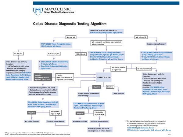
Small intestinal diseases are a common, though often overlooked cause of diarrhoeal illness. Fully 1% of the Caucasian population are affected by coeliac disease and a substantial portion of children living in poverty in the developing world are affected by environmental enteropathy. These are but two examples of the many diseases that cause mucosal injury to the primary digestive and absorptive organ in our body. While diarrhoea may be a common, though not universally seen symptom of small bowel mucosal disease, the consequent malabsorption can lead to substantial malnutrition and nutrient deficiencies. The small intestine, unlike the colon, has been relatively inaccessible, and systematic evaluation is often necessary to identify and treat small intestinal mucosal diseases that lead to diarrhoea. Immunodeficiency states, including HIV enteropathy, adult autoimmune enteropathy, drug-associated enteropathy, and tropical sprue continue to occur and require specific therapy. All patients with severe diarrhoea or diarrhoea associated with features suggestive of malabsorption may have a disease of the small intestinal mucosa that requires careful evaluation and targeted management.


Figures

Fig. 1
Fig. 1
Coeliac disease serology cascade based on total IgA measurement. [Courtesy of Melissa M. Snyder, PhD, Antibody immunology laboratory, Mayo Clinic].
Fig. 2
Fig. 2
Confirmation of the diagnosis of coeliac disease by using combined serology and histology. [Reprinted from Walker MM, Murray JA. An update in the diagnosis of coeliac disease. Histopathology 2011; 59:166–179].
Fig. 3
Fig. 3
A systematic approach to the evaluation of non-responsive coeliac disease. The outline of a systematic approach to the patient with non-responsive coeliac disease (NCD). LC=lymphocytic colitis. CC=collagenous colitis. GC=gluten contamination. RS=refractory sprue. PLE=protein-losing enteropathy. [Reprinted from Abdulkarim AS, Burgart LJ, See J, Murray JA. Etiology of nonresponsive coeliac disease: Results of a systematic approach. Am J Gastroenterol 2002; 97:2016–21].
Fig. 4
Fig. 4
Endoscopic appearance of villous atrophy in a patient with refractory coeliac disease. These findings are essentially indistinguishable from standard coeliac disease or indeed other causes of severe villous atrophy. [Reprinted from Rubio-Tapia A, Murray JA. Classification and management of refractory celiac disease. Gut 2010; 59:547–57].
Fig. 5
Fig. 5
Protocolized approach to the evaluation and management of patients with refractory coeliac disease. [Reprinted from Rubio-Tapia A, Kelly DG, Lahr BD, Dogan A, Wu T-T, Murray JA. Clinical staging and survival in refractory celiac disease: a single center experience. Gastroenterology 2009; 136:99–107].
Fig. 6
Fig. 6
Periodic-acid Schiff (PAS) positive macrophages and partial villous atrophy of the duodenum characteristic of Whipple’s disease (magnification: 10×).
Fig. 7
Fig. 7
Partial villous atrophy in a patient with tropical sprue is indistinguishable from mild gluten sensitive enteropathy on the basis of histology. Courtesy of Tsung-Teh Wu, M.D., Ph.D.
Fig. 8
Fig. 8
Photomicrograph of duodenal biopsies in a patient with autoimmune enteritis demonstrating near total villous atrophy. Note the dense lymphoplasma cellular infiltration of the lamina propria. (B) At a higher power, there is disruption of the brush border and neutrophilic infiltration of the crypts producing abscess. Note the absence of any surface lymphocytosis. [Reprinted from Akram S, Murray JA, Pardi DS, Alexander GL, Schaffner JA, Russo PA, Abraham SC. Adult autoimmune enteropathy: Mayo Clinic Rochester experience. Clin Gastroenterol Hepatol 2007; 5:1282–90].
Fig. 9
Fig. 9
This is an indirect immunofluorescence assay utilizing the serum from a patient with autoimmune enteropathy. The substrate used is small intestine. The pattern is a linear fluorescence pattern of the brush border of the enterocytes. Magnification: 300×. [Reprinted from Akram S, Murray JA, Pardi DS, Alexander GL, Schaffner JA, Russo PA, Abraham SC. Adult autoimmune enteropathy: Mayo Clinic Rochester experience. Clin Gastroenterol Hepatol 2007; 5:1282–90].
Fig. 10
Fig. 10
These photomicrographs illustrate total villous atrophy and features that are inherently similar to that of coeliac disease with crypt hyperplasia, mixed inflammatory infiltration involving the lamina propria, and a clear lucent area under the surface epithelium. (B) A trichrome stain highlights the collagen layer (arrows). Original magnification: 200×. [Reprinted from Rubio-Tapia A, Talley NJ, Gurudu SR, Wu TT, Murray JA. Gluten-free diet and steroid treatment are effective therapy for most patients with collagenous sprue. Clin Gastroenterol Hepatol 2010; 8:344–9].
Fig. 11
Fig. 11
Collagenous sprue represents a common pathologic endpoint to many particular triggers, which may consist of other primary inflammatory conditions, immune deficiencies, environmental exposures, particularly medications. [Reprinted from Rubio-Tapia A, Talley NJ, Gurudu SR, Wu TT, Murray JA. Gluten-free diet and steroid treatment are effective therapy for most patients with collagenous sprue. Clin Gastroenterol Hepatol 2010; 8:344–9].
Fig. 12
Fig. 12
Immunoglubulinemic sprue is a common gastrointestinal manifestation of combined variable immunoglobulin deficiency (CVID) (A; magnification: 4×). A distinguishing feature of this enteropathy is the absence of plasma cells (B; magnification 20×). Courtesy of Tsung-Teh Wu, M.D., Ph.D.

Similar articles

Cited by

References

    1. Rubio-Tapia A, Ludvigsson JF, Brantner TL, Murray JA, Everhart JE. The prevalence of coeliac disease in the United States. Am J Gastroenterol Oct. 2012;107(10):1538–44. - PubMed
    1. Rashtak S, Murray JA. Coeliac disease in the elderly. Gastroenterol Clin North Am. 2009 Sep;38(3):433–46. - PMC - PubMed
    1. Green PH, Cellier C. Celiac disease. N Engl J Med. 2007 Oct 25;357(17):1731–43. - PubMed
    1. Rampertab SD, Pooran N, Brar P, Singh P, Green PH. Trends in the presentation of celiac disease. Am J Med. 2006 Apr;119(4):e9–14. (355) - PubMed
    1. Jamma S, Rubio-Tapia A, Kelly CP, Murray J, Najarian R, Sheth S, et al. Celiac crisis is a rare but serious complication of celiac disease in adults. Clin Gastroenterol Hepatol. 2010 Jul;8(7):587–90. - PMC - PubMed

Publication types

MeSH terms

Substances

Supplementary concepts