Review
. 2013 Dec;63(7):688-95.
doi: 10.1016/j.neuint.2013.01.028. Epub 2013 Feb 4.

Glial adenosine kinase--a neuropathological marker of the epileptic brain

Affiliations
Review

Glial adenosine kinase--a neuropathological marker of the epileptic brain

Eleonora Aronica et al. Neurochem Int. 2013 Dec.

Abstract

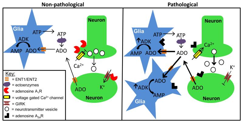
Experimental research over the past decade has supported the critical role of astrocytes activated by different types of injury and the pathophysiological processes that underlie the development of epilepsy. In both experimental and human epileptic tissues astrocytes undergo complex changes in their physiological properties, which can alter glio-neuronal communication, contributing to seizure precipitation and recurrence. In this context, understanding which of the molecular mechanisms are crucially involved in the regulation of glio-neuronal interactions under pathological conditions associated with seizure development is important to get more insight into the role of astrocytes in epilepsy. This article reviews current knowledge regarding the role of glial adenosine kinase as a neuropathological marker of the epileptic brain. Both experimental findings in clinically relevant models, as well as observations in drug-resistant human epilepsies will be discussed, highlighting the link between astrogliosis, dysfunction of adenosine homeostasis and seizure generation and therefore suggesting new strategies for targeting astrocyte-mediated epileptogenesis.

Keywords: ADK; Astrocytes; Epilepsy; Human; Rodents.


Figures

Figure 1
Figure 1. Astrocyte based ADK modulates network excitation by controlling the extracellular adenosine tone
Non-pathological state (left panel): Extracellular adenosine (ADO) arises from two sources including: (i) Ca2+ mediated release of ATP from astrocytes followed by catabolism to ADO through a series of ectoenzymes that include nucleoside triphosphate diphosphohydrolases, ectonucleotide pyrophosphatase/phosphodiesterases and ecto-5′-nucleotidases; and (ii) Direct ADO release into the extracellular space upon postsynaptic stimulation of neurons. Once in the extracellular space ADO inhibits neuron excitation through activation of A1Rs localized to the pre- and postsynaptic neuron membrane. On the presynaptic neuron, A1R activation inhibits Ca2+ dependent vesicular release of excitatory neurotransmitters by inhibiting P/Q- and N-type voltage gated Ca2+ channels; while on the postsynaptic neuron A1R hyperpolarizes the cell by activating G-protein coupled inwardly rectifying K+ channels (GIRKS). ADO is cleared from the extracellular space by passive propagation through concentrative and equilibrative transporters (ENT1/ENT2) on the astrocyte membrane. Once in the astrocyte cytoplasm ADO is metabolized into AMP by ADK, which sets the ambient ADO tone. Pathological state (right panel): Sustained neuronal excitation, as observed in epilepsy, induces a shift in adenosine receptor expression levels with A1R being superseded by A2AR. As a consequence, there is a loss of A1R activity, which translates to increased network excitability due to increased Ca2+ dependent vesicular release of excitatory neurotransmitters and attenuated GIRK mediated hyperpolarization. Furthermore, A2AR activation causes an increase in astrogliosis that is accompanied by increased ADK expression and activity. Pathological levels of ADK will drive ADO influx and metabolism; thereby, decreasing the extracellular ADO tone.
Figure 2
Figure 2. Increased ADK expression in a rodent model for mesial temporal lobe epilepsy
A,B ADK immunohistochemistry images from the contra- and ipsilateral hemisphere of a C57BL/6 mouse 10 weeks following intrahippocampal KA injection. A single unilateral injection of KA (400 ng/100 nL) into the CA1 subregion (coordinates relative to Bregma AP: −2.18; ML: −1.8, DV: −1.7) induces focal astrogliosis and associated ADK upregulation within the ipsilateral hippocampus (panel B), compared to the non-injured contralateral hemisphere (panel A). C,D High magnification images of the regions demarcated by boxes in panels A and B. Note that in the non-injured hemisphere (panel C) the nuclear isoform of ADK is predominantly expressed, while in the injured hemisphere there is a robust increase in cytoplasmic ADK expression within astrocytes (arrows, panel D).

References

    1. Abbracchio MP, Ceruti S. P1 receptors and cytokine secretion. Purinergic Signal. 2007;3:13–25. - PMC - PubMed
    1. Abbracchio MP, Rainaldi G, Giammarioli AM, Ceruti S, Brambilla R, Cattabeni F, Barbieri D, Franceschi C, Jacobson KA, Malorni W. The A3 adenosine receptor mediates cell spreading, reorganization of actin cytoskeleton, and distribution of Bcl-XL: studies in human astroglioma cells. Biochem Biophys Res Commun. 1997;241:297–304. - PMC - PubMed
    1. Aden U, O’Connor WT, Berman RF. Changes in purine levels and adenosine receptors in kindled seizures in the rat. Neuroreport. 2004;15:1585–1589. - PubMed
    1. Alanko L, Porkka-Heiskanen T, Soinila S. Localization of equilibrative nucleoside transporters in the rat brain. J Chem Neuroanat. 2006;31:162–268. - PubMed
    1. Albrecht-Kupper BE, Leineweber K, Nell PG. Partial adenosine A1 receptor agonists for cardiovascular therapies. Purinergic Signal. 2012;8:91–99. - PMC - PubMed

Web Reference

    1. Allen Brain Atlas mouse A1R expression profile. http://mouse.brainmap.org/search/show?page_num=0&page_size=20&no_paging=....

Publication types