Review
. 2013 Mar;36(2):339-55.
doi: 10.1007/s10545-013-9586-2. Epub 2013 Feb 6.

Spinal involvement in mucopolysaccharidosis IVA (Morquio-Brailsford or Morquio A syndrome): presentation, diagnosis and management

Affiliations
Review

Spinal involvement in mucopolysaccharidosis IVA (Morquio-Brailsford or Morquio A syndrome): presentation, diagnosis and management

Guirish A Solanki et al. J Inherit Metab Dis. 2013 Mar.

Abstract

Mucopolysaccharidosis IVA (MPS IVA), also known as Morquio-Brailsford or Morquio A syndrome, is a lysosomal storage disorder caused by a deficiency of the enzyme N-acetyl-galactosamine-6-sulphate sulphatase (GALNS). MPS IVA is multisystemic but manifests primarily as a progressive skeletal dysplasia. Spinal involvement is a major cause of morbidity and mortality in MPS IVA. Early diagnosis and timely treatment of problems involving the spine are critical in preventing or arresting neurological deterioration and loss of function. This review details the spinal manifestations of MPS IVA and describes the tools used to diagnose and monitor spinal involvement. The relative utility of radiography, computed tomography (CT) and magnetic resonance imaging (MRI) for the evaluation of cervical spine instability, stenosis, and cord compression is discussed. Surgical interventions, anaesthetic considerations, and the use of neurophysiological monitoring during procedures performed under general anaesthesia are reviewed. Recommendations for regular radiological imaging and neurologic assessments are presented, and the need for a more standardized approach for evaluating and managing spinal involvement in MPS IVA is addressed.


Conflict of interest statement

G. A. Solanki, K. W. Martin and M. C. Theroux have each received speaker’s honorarium and travel support from BioMarin Pharmaceutical Inc. (BioMarin). C. Lampe has received speaker’s fees, consulting fees, travel support and research grants from BioMarin. K. K. White has received honoraria, research grants and travel support from BioMarin, and is an investigator in MPS IVA clinical trials. R. Shediac is an employee and shareholder of BioMarin. C.G. Lampe and W. G. Mackenzie have received travel support from BioMarin. M. Beck has received speaker’s fees, consulting fees, travel support and unrestricted research grants from Biomarin. C. J. Hendriksz has received consulting fees, symposium support for himself and team personnel, and research grants from BioMarin, and has also been an investigator in MPSIVA clinical trials. P. R. Harmatz has provided consulting support to BioMarin, has received research grants, speaker’s honorarium and travel support from BioMarin, and is an investigator in MPS IVA clinical trials.

Figures
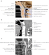
Fig. 1
Fig. 1
Normal anatomy of the craniocervical junction. a Sagittal midline graphic depicts the normal articulations and ligamentous anatomy of the craniocervical region. b Sagittal midline CT reconstruction shows the bony and ligamentous structures at the craniocervical junction. c Sagittal T2-weighted MR image of the craniocervical junction shows the bony and ligamentous components and the cervical spinal cord. Images reproduced with permission from Amirsys Publishing, Inc. (Harnsberger et al 2006)
Fig. 2
Fig. 2
Spine radiographs demonstrating the manifestations of MPS IVA. a Lateral view of the cervical spine shows a cone-shaped hypoplastic dens, which may be associated with upper cervical spine instability. The unossified cartilage cap is inapparent. The posterior arch of C1 is small, but thickened, resulting in spinal stenosis. b Typical thoracolumbar spine changes including platyspondyly, anterior beaking, thoracolumbar kyphosis, posterior vertebral scalloping, and broadened ribs
Fig. 3
Fig. 3
Assessing cervical instability. Cervical flexion-extension lateral radiographs in (a) can be difficult to interpret. The pre-dental space is obscured by delayed ossification of the dens and superimposition of the mastoid processes (arrow). Measuring the spinal canal at C1-2 between the base of the odontoid process of C2 and the posterior arch of C1 is preferred (white lines). Flexion-extension CT with sagittal reformation and soft-tissue filtration in (b) shows the cone-shaped dens, a thickened cruciate ligament, and small thick cartilaginous posterior arch of C1 (arrow). With flexion, there is narrowing of the canal between the body of C2 and the cartilaginous posterior arch of C1 (white lines). T2 TSE sagittal flexion-extension MR has the advantage of imaging the spinal canal and cord directly. As shown in (c), there is spinal stenosis and flexion instability sufficient to compress the cervical cord at C1-2 (arrow)
Fig. 4
Fig. 4
Initial MRI studies should include screening of the whole spine. In (a) T2 TSE sagittal MR shows intermediate signal in flattened vertebral bodies (short arrow) separated by hypointense bulging discs (long arrow). Typical cervical lordosis, cervicothoracic and thoracolumbar kyphosis, and multiple areas of spinal stenosis are present. Smaller field-of-view imaging optionally characterizes areas of stenosis as shown by T2 TSE sagittal MR at C1-2. In (b) delayed ossification of the odontoid process of C2 and the small thickened posterior arch of C1 are present with dorsal cord compression (arrow). In (c) bFFE sagittal MR of the thoracolumbar kyphosis shows a bulging disc at T11 causing ventral compression of the conus medullaris (arrow). Dephasing of accelerated CSF causing signal loss is present dorsal to the cord at the stenosis
Fig. 5
Fig. 5
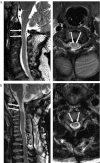
Myelomalacia is best depicted by T2-weighted MR obtained in the sagittal and axial planes where an increase in T2 signal, coupled with volume loss in regions of cord compression, is diagnostic of myelomalacia. When T2 hyperintensity is present without atrophy, the differential diagnosis would include edema due to contusion or microvascular injury. These changes may be more apparent following surgical decompression. In (a), there is chronic T2 hyperintensity and little or no atrophy, indicating mild-to-moderate injury. With more severe cord injury, as shown in (b), there is T2 hyperintensity and focal atrophy. Postoperative T2 TSE axial and sagittal MR images show central grey matter involvement in both examples (arrows)

References

    1. Ahmed R, Traynelis VC, Menezes AH. Fusions at the craniovertebral junction. Childs Nerv Syst. 2008;24:1209–1224. doi: 10.1007/s00381-008-0607-7. - DOI - PubMed
    1. Ain MC, Chaichana KL, Schkrohowsky JG. Retrospective study of cervical arthrodesis in patients with various types of skeletal dysplasia. Spine (Phila Pa 1976) 2006;31:E169–174. doi: 10.1097/01.brs.0000202758.61848.61. - DOI - PubMed
    1. Ashraf J, Crockard HA, Ransford AO, Stevens JM. Transoral decompression and posterior stabilisation in Morquio’s disease. Arch Dis Child. 1991;66:1318–1321. doi: 10.1136/adc.66.11.1318. - DOI - PMC - PubMed
    1. Beighton P, Craig J. Atlanto-axial subluxation in the Morquio syndrome. Report of a case. J Bone Joint Surg Br. 1973;55:478–481. - PubMed
    1. Belani KG, Krivit W, Carpenter BL, et al. Children with mucopolysaccharidosis: perioperative care, morbidity, mortality, and new findings. J Pediatr Surg. 1993;28:403–408. doi: 10.1016/0022-3468(93)90240-L. - DOI - PubMed

MeSH terms