. 2013 Feb;69(Pt 2):266-75.
doi: 10.1107/S0907444912045520. Epub 2013 Jan 19.

Identification of quercitrin as an inhibitor of the p90 S6 ribosomal kinase (RSK): structure of its complex with the N-terminal domain of RSK2 at 1.8 Å resolution

Affiliations

Identification of quercitrin as an inhibitor of the p90 S6 ribosomal kinase (RSK): structure of its complex with the N-terminal domain of RSK2 at 1.8 Å resolution

Urszula Derewenda et al. Acta Crystallogr D Biol Crystallogr. 2013 Feb.

Abstract

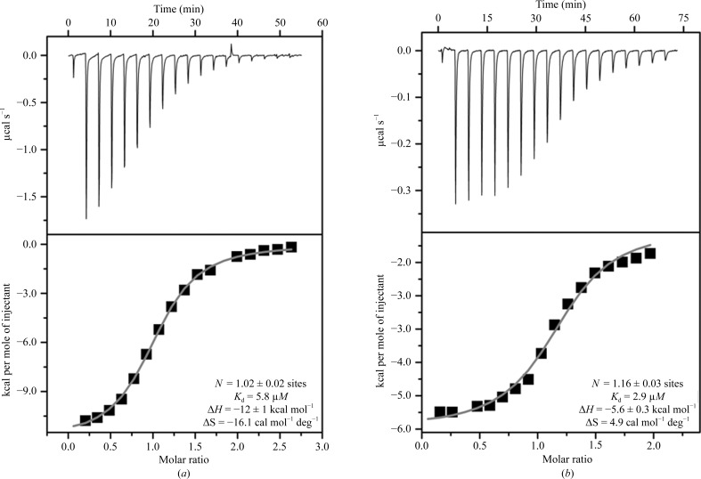
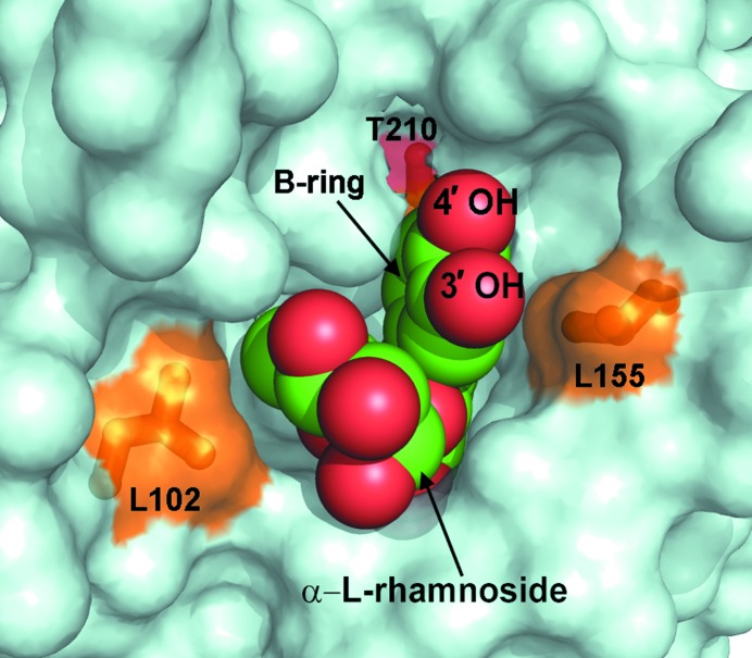
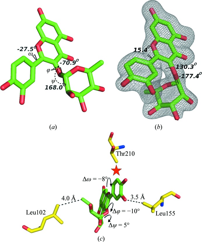
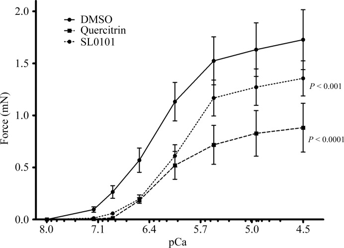
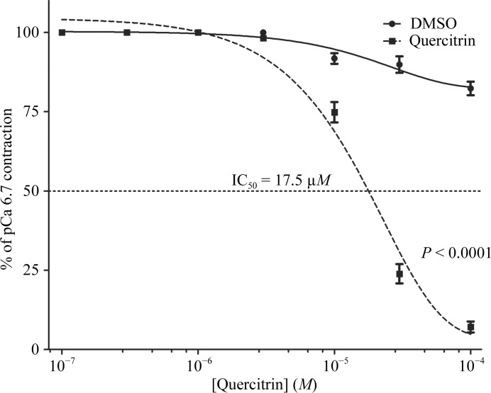
Members of the RSK family of kinases constitute attractive targets for drug design, but a lack of structural information regarding the mechanism of selective inhibitors impedes progress in this field. The crystal structure of the N-terminal kinase domain (residues 45-346) of mouse RSK2, or RSK2(NTKD), has recently been described in complex with one of only two known selective inhibitors, a rare naturally occurring flavonol glycoside, kaempferol 3-O-(3'',4''-di-O-acetyl-α-L-rhamnopyranoside), known as SL0101. Based on this structure, it was hypothesized that quercitrin (quercetin 3-O-α-L-rhamnopyranoside), a related but ubiquitous and inexpensive compound, might also act as an RSK inhibitor. Here, it is demonstrated that quercitrin binds to RSK2(NTKD) with a dissociation constant (K(d)) of 5.8 µM as determined by isothermal titration calorimetry, and a crystal structure of the binary complex at 1.8 Å resolution is reported. The crystal structure reveals a very similar mode of binding to that recently reported for SL0101. Closer inspection shows a number of small but significant differences that explain the slightly higher K(d) for quercitrin compared with SL0101. It is also shown that quercitrin can effectively substitute for SL0101 in a biological assay, in which it significantly suppresses the contractile force in rabbit pulmonary artery smooth muscle in response to Ca(2+).

Keywords: SL0101; flavonol glycosides; inhibitors; protein kinases; quercitrin.


Figures

Figure 1
Figure 1
The chemical structures of the inhibitor SL0101 and its analogue quercitrin. The conformational dihedral angles ω, ϕ and ψ are also shown, together with the canonical numbering scheme of the whole molecule.
Figure 2
Figure 2
Thermodynamics of the binding of quercitrin (a) and SL0101 (b) to the N-terminal domain of mouse RSK2. The upper panels show raw titration data, while the lower panels show integrated titration curves and the final values of the determined thermodynamic parameters. The data for SL0101 are included for comparison and were originally determined and published in Utepbergenov et al. (2012 ▶).
Figure 3
Figure 3
Overview of the crystal structure of the complex of quercitrin with the N-­terminal kinase domain of mouse RSK2. All main secondary-structure elements, as well as the N- and C-lobes, are labeled. The Gly-rich loop is colored orange, the β-strands of the RSK-specific N-lobe sheet are shown in different shades of blue and the catalytic loop is colored purple. The hinge motif and the bound quercitrin are labeled.
Figure 4
Figure 4
A comparison of the binding modes of adenine and flavonols to protein kinases. (a) The binding mode of the adenine moiety of AMPPNP to the hinge region of mRSK2NTKD (PDB entry 3g51; Malakhova et al., 2009 ▶). (b) The binding mode of quercetin to the hinge of protein kinase 17B (PDB entry 3lm5; Structural Genomics Consortium, unpublished work). (c) The binding mode of SL0101 to the hinge of mRSK2NTKD (PDB entry 3ubd; Utepbergenov et al., 2012 ▶). The binding mode of quercitrin reported in this study is identical to that of SL0101.
Figure 5
Figure 5
The quercitrin molecule nested in the binding pocket between the N- and C-lobes of mRSK2NTKD. Quercitrin is shown using van der Waals spheres (oxygen in red and carbon in green); the protein is depicted as a surface representation. The three key residues making close contacts with quercitrin (and also depicted in Fig. 6 ▶) are shown as sticks and are colored and labeled.
Figure 6
Figure 6
The structure of quercitrin unbound and bound to mRSK2NTKD. (a) Free quercitrin (CCDC code 698519), shown with H atoms not visualized and with the three dihedral conformational angles shown and labeled. (b) The conformation of quercitrin bound to mRSK2NTKD, shown along with an OMIT electron-density map contoured at 2σ; the values of the dihedral angles are shown. (c) A diagrammatic representation of the differences in the stereochemistry of bound quercitrin compared with bound SL0101.
Figure 7
Figure 7
The RSK2 inhibitors quercitrin and SL0101 decrease the maximal force at each intracellular Ca2+ concentration (pCa). The significance of the difference between curves was determined using two-way analysis of variance. Corresponding column factor P values are shown on each graph; n = 4–9.
Figure 8
Figure 8
A dose-response curve for quercitrin in α-toxin-permeabilized rabbit pulmonary artery smooth muscle. The significance of the difference between curves was determined using two-way analysis of variance. P < 0.0001 versus diluent control; n = 3–4.

Similar articles

Cited by

References

    1. Adams, P. D. et al. (2010). Acta Cryst. D66, 213–221.
    1. Anjum, R. & Blenis, J. (2008). Nature Rev. Mol. Cell Biol. 9, 747–758. - PubMed
    1. Bain, J., Plater, L., Elliott, M., Shpiro, N., Hastie, C. J., McLauchlan, H., Klevernic, I., Arthur, J. S., Alessi, D. R. & Cohen, P. (2007). Biochem. J. 408, 297–315. - PMC - PubMed
    1. Boly, R., Gras, T., Lamkami, T., Guissou, P., Serteyn, D., Kiss, R. & Dubois, J. (2011). Int. J. Oncol. 38, 833–842. - PubMed
    1. Calderón-Montaño, J. M., Burgos-Morón, E., Pérez-Guerrero, C. & López-Lázaro, M. (2011). Mini Rev. Med. Chem. 11, 298–344. - PubMed

Publication types

MeSH terms