. 2013:4:1431.
doi: 10.1038/ncomms2441.

The microbiome extends to subepidermal compartments of normal skin

Affiliations

The microbiome extends to subepidermal compartments of normal skin

Teruaki Nakatsuji et al. Nat Commun. 2013.

Abstract

Commensal microbes on the skin surface influence the behaviour of cells below the epidermis. We hypothesized that bacteria or their products exist below the surface epithelium and thus permit physical interaction between microbes and dermal cells. Here to test this hypothesis, we employed multiple independent detection techniques for bacteria including quantitative PCR, Gram staining, immunofluorescence and in situ hybridization. Bacteria were consistently detectable within the dermis and dermal adipose of normal human skin. Sequencing of DNA from dermis and dermal adipose tissue identified bacterial 16S ribosomal RNA reflective of a diverse and partially distinct microbial community in each skin compartment. These results show the microbiota extends within the dermis, therefore, enabling physical contact between bacteria and various cells below the basement membrane. These observations show that normal commensal bacterial communities directly communicate with the host in a tissue previously thought to be sterile.


Figures
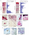
Figure 1
Figure 1. Bacterial components are detectable deep in normal human skin
(a-b) Total DNA was extracted from sequential 30 or 50 μm horizontal sections of 6 mm punch biopsies of human facial (a) or palm skin (b). 16S rRNA gene was quantified with real-time qPCR using universal 16S rRNA primers. Relative abundance of 16S rRNA gene was calculated by the ΔCt method. H&E staining of each skin biopsy is shown in the same scale as the Y-axis of the graph. Arrow indicates data from the DNA sample prepared from a non-tissue control (NTC) that represents a section of OCT embedding compound devoid of tissue and processed with all reagents used for amplification of tissue sections. See Supplementary Fig. S1 for additional biopsy data from different donors. (c) Gram staining of frozen sections from normal human facial skin. See Supplementary Fig. S4 for additional biopsy data from different donors. (d) To examine if bacteria are associated with CD11c+ cells, normal human facial skin was Gram stained (purple/ red: arrow), and then stained with anti-CD11c monoclonal IgG (brown: arrow head). Bar= 20μm.
Figure 2
Figure 2. Detection of microbes in normal human skin by immunostaining and in situ hybridization
(a-d) Normal human facial skin was stained with anti-S. epidermidis monoclonal IgG (a and b) or isotype IgG (c and d). The broken line depicts the basement membrane of the epidermis. (e-h) Normal human facial skin was stained with anti-Pseudomonas spp. (green) and anti-keratin 14 (K14) (red) (e and g), or isotype control IgGs (f and h). (i-j) LPS was detectable in the reticular dermis of facial skin sections stained with anti-LPS (green) and was associated with collagen fibers detected by anti-collagen I staining (blue). Phagocytic cells stained with anti-CD11c IgG (red) were undetectable (i). Control IgGs for LPS, collagen I and CD11c were negative in adjacent sections (j). (k-l) Bacterial 16S rRNA was detectable in normal human facial skin sections hybridized with an Alexa Fluor488-labeled EUB338 probe. Specific hybridization was detected in dermal adipose tissue (k). No signal was seen in adjacent sections hybridized with a nonsense control probe (nonEUB338) (l). See Supplementary Fig. S5 for additional biopsy data from different donors. Bar= 20μm, if not indicated.
Figure 3
Figure 3. The subcutaneous microbiome is diverse
(a) The profile of the subcutaneous microbiome at the class level derived from pyrosequencing analysis of DNA encoding the 16S rRNA gene. Relative abundance of bacterial classes associated with each skin compartment is shown for each skin biopsy. (b) PCoA plot analysis displaying similarity of composition of bacterial 16S rRNA gene sequences contained in each cutaneous compartment. Each symbol corresponds to the sample of epidermis(■), follicle(▲), dermis(•) and adipose tissue (×). The biopsy number is shown in different color and identical to the data in Fig. 3a.
Figure 4
Figure 4. Measurement of abundance of bacterial DNAs in subepidermal compartments
(a-c) Abundance of universal bacterial 16S rRNA gene (a), P. acnes-specific 16S rRNA gene (b), S. epidermidis SodA gene (c), or Pseudomonas-specific 16S rRNA gene (d) was measured by real-time qPCR. Relative abundance of DNA for 16S rRNA gene was calculated with a ΔCt method. P. acnes and S. epidermidis were quantified by comparison to known colony forming units (CFUs) of these organisms. Pseudomonas species was quantified by comparison to known CFUs of Pseudomonas aeruginosa. Skin compartments including epidermis and stratum corneum, follicle, dermis, and subcutaneous adipose tissue was excised by LCM. Non-tissue control (NTC) processed from embedding material adjacent to the tissue sample, and sterile skeletal muscle biopsy tissue, are also shown as negative controls to detect contamination. Each point represents a separate individual skin biopsy. The biopsy number is shown in different color and identical to the data in Fig. 3a. The skeletal muscle specimens shown were obtained from five different donors.

References

    1. Wilhelm KP, Cua AB, Maibach HI. Skin aging. Effect on transepidermal water loss, stratum corneum hydration, skin surface pH, and casual sebum content. Arch Dermatol. 1991;127:1806–1809. - PubMed
    1. Ali RS, et al. Expression of the peptide antibiotics human beta defensin-1 and human beta defensin-2 in normal human skin. J Invest Dermatol. 2001;117:106–111. - PubMed
    1. Grice EA, et al. Topographical and temporal diversity of the human skin microbiome. Science. 2009;324:1190–1192. - PMC - PubMed
    1. Capone KA, Dowd SE, Stamatas GN, Nikolovski J. Diversity of the human skin microbiome early in life. J Invest Dermatol. 2011;131:2026–2032. - PMC - PubMed
    1. Costello EK, et al. Bacterial community variation in human body habitats across space and time. Science. 2009;326:1694–1697. - PMC - PubMed

Publication types